New biomedical imaging technology could enhance pathologists’ ability to examine tissue samples via fluorescence microscopy
Scientists at Harvard University’s Wyss Institute for Biologically Inspired Engineering have developed a new DNA, barcoding technique. The fluorescence microscopy approach has significant implications for the imaging community.
Beyond imaging, however, pathologists will be able to use this same technology when evaluating tissue specimens.
The new method could enable simultaneous imaging of many different types of molecules in a single cell, according to Peng Yin, Ph.D., Associate Professor of Systems Biology at Harvard Medical School and Core Faculty Member at Wyss Institute. The developers expect the method to provide researchers with a richer, more accurate view of cell behavior than is possible using current techniques.
Pathologists Could Adopt DNA Barcoding for In Vitro Diagnostics
“We hope this new method will provide much-needed molecular tools for using fluorescence microscopy to study complex biological problems,” stated Yin, the study’s co-author, in a recent press release.
Using DNA Origami to Create Fluorescent Linear DNA Barcodes
The newly engineered DNA barcode harnesses the natural ability of DNA to self-assemble. The basis of the new technology is a process called DNA origami. This enables scientists to arrange colored dots, or fluorophores, into geometric patterns, or fluorescent linear DNA barcodes.

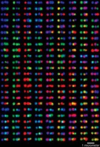
Researchers at Harvard’s Wyss Institute recently engineered a new DNA barcode. Labeled DNA samples appear as multi-colored barcodes under fluorescent light at certain wavelengths. Pathologists and clinical laboratory professionals will recognize the potential of this technology in the examination of tissue specimens. (Photo credit: Rick Groleau, Harvard University.)
These imaging probes translate a cell’s invisible biological information, such as proteins or RNA molecules, into detectable signals, noted a summary of Yin’s research on the Wyss website. These signals help researchers better understand the role of cell behavior in the onset and progression of disease.
New Barcode Could Offer a Virtually Unlimited Number of Styles
Scientists currently use fluorescence microscopy to pair fluorescent elements—the barcodes—with molecules they know will attach to the part of the cells they want to investigate. When they illuminate the sample, it triggers each kind of barcode to fluoresce at a particular wavelength of light, which indicates the location of the molecules of interest.
However, the multiplexing ability of fluorescence microscopy is limited by the number of spectrally distinguishable fluorophores, a story in Nature Chemistry explained. The barcodes that scientists currently use have only three or four colors available, such as red, blue, or green. And sometimes those colors blur. This limits the number of objects scientists have been able to study in a cell sample at one time.
Multiplex Capability with 216 Readable Color Combinations
Using the new method, Yin was able to demonstrate 216 color combinations resulting from attaching just three colors to a DNA nanotube, the press release stated. With the new barcode, the combinations are almost limitless. This will significantly advance the ability to fluoresce more cellular structure than previously possible.
DNA origami works by programming a long strand of DNA to self-assemble by folding in on itself, the release stated. Shorter strands, called staples, help it to create predetermined forms. Researchers then attach fluorescent molecules to the desired spots on the now more structurally complex DNA nanostructures. In this way, they use origami technology to generate a large pool of barcodes out of only a few fluorescent molecules.
“We can essentially use DNA joints to assemble the nanostructures into long rods, and we can modify the rods with fluorescence at different locations,” declared Yin. “We then use these tiny rods to arrange the fluorescent spots into colorful barcodes. Basically now using three or four colors, we can have hundreds of different barcodes,” he observed in a story in The Harvard Crimson.
A New Tool in the Cellular Imaging—and In Situ Examination—Toolbox
“[The technique] holds great promise for using the method to study cells in their native environments,” Yin observed. Additionally, the technique is low-cost, easy to do, and more robust compared to current methods, according to Yin.
Pathologists and clinical laboratory managers will recognize the range of potential of this new technology, from developing targeted drug-delivery mechanisms to improving the scope of cellular and molecular activities scientists are able to observe at a disease site.
—Pamela Scherer McLeod
Related Information:
Novel DNA Barcode Engineered: New Technology Could Launch Biomedical Imaging to Next Level
Researchers at Harvard’s Wyss Institute Engineer Novel DNA Barcode
Scientists Develop DNA Barcoding
A primer to scaffolded DNA origami
Engineered DNA barcode boosts fluorescence microscopy potential


