Second-generation device is self-powered, does not require a trained operator, and amplifies the fluorescence signal by 1,000-fold, enabling early detection of cancer
Pathologists will be interested to learn that Japanese researchers have developed a second-generation lab-on-a-chip that detects microRNA (miRNA) from a tiny sample volume in only 20 minutes! Their goal is to create a point-of-care device for early detection of cancer.
This is another example of how a variety of fast-developing technologies are being brought together to create diagnostic testing systems that have capabilities that challenge the clinical laboratory analyzers used in centralized medical laboratories.
Creating More Capabilities to Advance Clinical Laboratory Medicine
Scientists achieved this groundbreaking work at Japan’s RIKEN Advanced Science Institute. Their first generation device required too large of a sample volume to make the device practical and days to get an answer. The equipment also required trained personnel to operate it.
“Rapidness, simple operation, small required sample volume, and portability of the device are ideal advantages for point-of-care cancer diagnosis,” stated study co-authors Hideyuki Arata, Ph.D., Hiroshi Komatsun and Kazuo Hosokawa, Ph.D.. Their paper was published by PLOS ONE.
MiRNA Could Hold Key to Earlier Detection of Disease
MiRNAs are small, non-coding RNA molecules. They regulate gene expression in a wide range of biological processes, including development, cell proliferation, differentiation and cell death. MiRNAs have become increasingly important in determining disease diagnosis and prognosis, according to the researchers’ abstract.

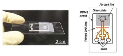
Researchers at Japan’s RIKEN Advanced Science Institute developed a self-powered microfluidic chip for cancer biomarker detection. A tiny sample and two fluorescence amplification reagents are added to the three inlet ports. Fluorescence in the main microfluidic channel indicates the presence of cancer biomarkers. This research is expected to lead to a point-of-care medical testing device that would support early detection of cancer and other diseases. It is a technology that could shift medical laboratory testing out of clinical laboratories and into physicians’ offices and other near-patient settings. (Image copyright Riken Institute.)
Concentration of certain miRNA in body fluids increases with the progression of diseases such as cancer and Alzheimer’s. Researchers hope, therefore, that these short RNA may hold the key to faster, more accurate diagnosis for diseases like these, stated a RIKEN press release.
“The development of an inexpensive and rapid point-of-care diagnostic test capable of spotting such early biomarkers of disease could therefore save many lives,” noted a RIKEN Research story about this work on the Instititue’s website.
Fluorescence Signal Amplification Improves Sensitivity One Thousand-fold
In their earlier research, the RIKEN team developed a microchip device which used the potential energy of degassed polydimethylsiloxane (PDMS) as a pumping technique, allowing the mechanical pump to draw reagents into a capture probe for analysis, according to a RIKEN press release. The technique eliminated the need for external power sources. However, it required a sample quantity too large for practical applications.
In the new study, the team used a signal amplification method called laminar flow-assisted dendritic amplification (LFDA). The LFDA technique allowed them to accurately detect microRNA from a sample of just 0.25 attomoles (10-18 mole, or 0.000 000 000 000 000 001).
The researchers dramatically boosted sensitivity in the new chip by adding a fluorescence amplification process. The process involves passing two amplification reagents over immobilized target microRNA. The reagents bind to immobilized miRNA to form tree-like dendritic structures. These amplify the fluorescence signal by up to 1,000 times. This amplification gives the device a level of sensitivity approaching that required for early detection of cancer.
POC Testing for Early-stage Cancer and Other Diseases
Pathologists and clinical laboratory administrators recognize the paradigm-shifting significance of low-cost point-of-care testing with the speed and sensitivity to diagnose early stage diseases. Such advances in diagnostic technology are especially important for remote and resource-limited areas of the world.
“[F]urther study might contribute to improvement of the healthcare environment, even in resource-poor environments of developing countries, and may have both an industrial and societal impact on global health,” the researchers wrote.
The next step for the team will be to further simplify the device by eliminating the need for a fluorescence microscope. This will involve replacing the fluorescent tags with some other form of marker.
“That is a very important direction for future development,” observed study author Hosokawa in the phys.org piece. “We are planning the use of different labeling materials instead of the fluorescent dye, such as gold particles, which would enable naked-eye detection,” he revealed. The team is also working to further improve the sensitivity of the technique.
Although in need of much refinement before it can be adapted and approved for clinical use, this new device shows how researchers are combining multiple technology developments to create diagnostic assay devices that signal a new paradigm for clinical laboratory testing.
—Karen Branz
Related Information:
Rapid cancer detection on a chip
New portable device enables RNA detection from ultra-small sample in only 20 minutes
DNA detection on a power-free microchip with laminar flow-assisted dendritic amplification


