Innovative and inexpensive technologies hold the promise of instantaneous diagnosis while transforming conventional clinical laboratory tools
“Point-of-care pathology” may not be that far away! The convergence of medical and information technologies, the falling cost of computing, and the growing availability of miniaturization technologies make it increasingly possible for pathologists and physicians to make informed, on-the-spot diagnostic decisions about patient care.
A new wave of imaging technologies—including pathology tools—is poised to transform the practice of medicine, reported a recent story in the New York Times. Some of these technologies make it possible for pathologists and other physicians to view individual cells in situ and in vivo.
The NY Times story highlighted multiple research and development initiatives that have the same goal: reduce the time get an answer for tissue diagnosis and bring diagnostic tests to the patient. Some technologies would engage pathologists. Others reflect developments in radiology and imaging.
Next-generation Endoscope Reduces Time between Tissue Sample and Examination
A microbiologist at Stanford University has created a variety of noninvasive imaging instruments, the NY Times story reported. His next-generation endoscopes allow pathologists to probe the esophagus, stomach and intestines looking for cancers. The instruments use a variety of optical and acoustical techniques to provide a three-dimensional view below the surface of the skin. The approach uses contrast agents to identify abnormalities.

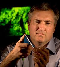
Pictured above is Christopher H. Contag, Ph.D., Professor of Microbiology and Immunology at Stanford University School of Medicine. He is developing imaging tools that would allow the in vivo study of cellular and molecular biology. Such tools bring closer to the day pathology when point–of-care diagnostics becomes feasible. (Photo copyright Stanford University)
“The imaging tools that we develop are sensitive, image over a range of scales from micro- to macroscopic, and are well-suited for the in vivo study of cellular and molecular biology,” stated Christopher H. Contag, Ph. D., Professor of Microbiology and Immunology, and Professor (by courtesy) of Radiology at Stanford.
Additionally, Contag is working on a new generation of injectable molecular biomarkers that would attach to lesions. This would enable physicians to get direct answers about disease on a cell-by-cell basis, reported the NY Times. Contag demonstrated with an image of a digital sample on a computer screen. Simply based on the dark and light sections, he noted, a physician could determine, “That’s cancer; that’s normal.”
Machine Imaging and Computation To Transform Conventional Lab Tools
Meanwhile, scientists at Columbia University Medical Center have developed pattern recognition software that can speed up the pathologist’s work, the NY Times reported. The software, known as nSPEC, can do in just 15 minutes what could take a full day using traditional pathology methods, according to Matthew Putman, Ph. D., Adjunct Assistant Professor and Adjunct Associate Research Scientist at The Fu Foundation School of Engineering and Applied Sciences at Columbia.
This isn’t a case of replacing the pathologists, the NY Times writer pointed out. It is a case of replacing the centralized multimillion-dollar systems that now do the preliminary screening used to look for abnormalities.
Advances in computation are rapidly transforming other traditional imaging technologies. Some examples include innovations being developed by Royal Philips Electronics (NYSE: PHG; AEX: PHI). Philips has used computation extensively in various technology innovations, the NY Times stated. Examples include:
- Three-dimensional transesophageal echocardiography (3-D TEE), an advanced ultrasound system inserted through the patient’s mouth into the esophagus. It creates a “stunning high-resolution 3-D video of the beating heart.”
- HeartNagigator, a system which provides a 3-D map for cardiologists, enabling the routine implantation of replacement heart valves by catheter.
- Magnetic particle imaging (M.P.I.), with higher resolution and faster imaging, holds promise for tumor diagnosis and coronary assessments. The technology could play a crucial role in surgical procedures that now rely on CT and PET scans. M.P.I.
Forward-looking pathologists and clinical laboratory administrators will recognize these advancements in pathology and imaging tools as an opportunity to increase accuracy and precision of diagnoses.
At the same time, they will want to assure that their medical laboratories are prepared to integrate new technologies into their workflow in a way that increases lab productivity.
Related Information:
Laboratories Seek New Ways to Take a Look Inside
Fewer Biopsies Go to Pathology Labs When Gastroenterologists Use New Miniature Microscope
Digital Pathology Should Leapfrog Digital Radiology’s Adoption Timeline
Helping Pathologists Use New Technology to Identify and Classify Cancer-Related Cells Research


