VCU scientists used the technique to measure mutations associated with acute myeloid leukemia, potentially offering an attractive alternative to DNA sequencing
More accurate but less-costly cancer diagnostics are the Holy Grail of cancer research. Now, research scientists at Virginia Commonwealth University (VCU) say they have developed a clinical laboratory diagnostic technique that could be far cheaper and more capable than standard DNA sequencing in diagnosing some diseases. Their method combines digital polymerase chain reaction (dPCR) technology with high-speed atomic force microscopy (HS-AFM) to generate nanoscale-resolution images of DNA.
The technique allows the researchers to measure polymorphisms—variations in gene lengths—that are associated with many cancers and neurological diseases. The VCU scientists say the new technique costs less than $1 to scan each dPCR reaction.
The researchers used the technique to measure and quantify polymorphisms associated with mutations in the FLT3 gene. Cancer researchers have linked these mutations, known as internal tandem duplications (ITDs), to a poor prognosis of acute myeloid leukemia (AML) and a more aggressive form of the disease, Nature Leukemia noted in “Targeting FLT3 Mutations in AML: Review of Current Knowledge and Evidence.”
“We chose to focus on FLT3 mutations because they are difficult to [diagnose], and the standard assay is limited in capability,” said physicist Jason Reed, PhD, Assistant Professor in the Virginia Commonwealth University Department of Physics, in a VCU press release.
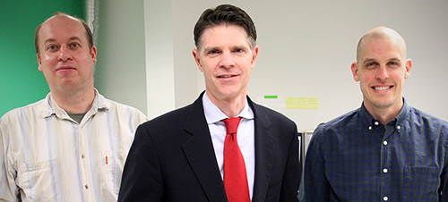
Reed is an expert in nanotechnology as it relates to biology and medicine. He led a team that included other researchers in VCU’s physics department as well as physicians from VCU Massey Cancer Center and the Department of Internal Medicine at VCU School of Medicine.

Validating the Clinical Laboratory Test
The physicists worked with two VCU physicians—hematologist/oncologist Amir Toor, MD, and hematopathologist Alden Chesney, MD—to compare the imaging technique to the LeukoStrat CDx FLT3 Mutation Assay, which they described as the “current gold standard test” for diagnosing FLT3 gene mutations.
The researchers said their technique matched the results of the LeukoStrat test in diagnosing the mutations. But unlike that test, the new technique also can measure variant allele frequency (VAL). This “can show whether the mutation is inherited and allows the detection of mutations that could potentially be missed by the current test,” states the VCU press release.
The VCU researchers published their findings in ACS Nano, a journal of the American Chemical Society (ACS), titled, “Digital Polymerase Chain Reaction Paired with High-Speed Atomic Force Microscopy for Quantitation and Length Analysis of DNA Length Polymorphisms.” They also presented their findings at the annual meetings of the Association of Molecular Pathology (AMP) and American Society of Hematology (ASH).
“We plan to continue developing and testing this technology in other diseases involving DNA structural mutations,” Reed said. “We hope it can be a powerful and cost-effective tool for doctors around the world treating cancer and other devastating diseases driven by DNA mutations.”
How the New Diagnostic Technique Works
Sean Koebley, PhD, Postdoctoral Fellow at Virginia Commonwealth University and another member of the VCU research team, described the new diagnostic technique in a video produced for the ASH and AMP meetings.
“In our approach we first used digital PCR, in which a mixed sample is diluted to less than one target molecule per aliquot and the aliquots are amplified to yield homogeneous populations of amplicons,” he said. “Then, we deposited each population onto an atomically-flat partitioned surface.”
The VCU researchers “scanned each partition with high-speed atomic force microscopy, in which an extremely sharp tip is rastered across the surface, returning a 3D map of the surface with nanoscale resolution,” he said. “We wrote code that traced the length of each imaged DNA molecule, and the distribution of lengths was used to determine whether the aliquot was a wild type [unmutated] or variant.”
In Diagnostics World, Reed said the method “doesn’t really have any more complexity than a PCR assay itself. It can easily be done by most lab technicians.”
Earlier Research
A VCU press release from 2017 noted that Reed’s research team had developed technology that uses optical lasers (similar to those in a DVD player) to accelerate the scanning. The researchers previously published a study about the technique in Nature Communications, and a patent is currently pending.
“DNA sequencing is a powerful tool, but it is still quite expensive and has several technological and functional limitations that make it difficult to map large areas of the genome efficiently and accurately,” Reed said in the 2017 VCU press release. “Our approach bridges the gap between DNA sequencing and other physical mapping techniques that lack resolution. It can be used as a stand-alone method or it can complement DNA sequencing by reducing complexity and error when piecing together the small bits of genome analyzed during the sequencing process.”
Using CRISPR technology, the team also developed what they described as a “chemical barcoding solution,” placing markers on DNA molecules to identify genetic mutations.
New DNA Clinical Laboratory Testing?
Cancer diagnostics are constantly evolving and improving. It is not clear how long it will be before VCU’s new technique will reach clinical laboratories that perform DNA testing, if at all. But VCU’s new technique is intriguing, and should it prove viable for clinical diagnostic use it could revolutionize cancer diagnosis. It is a development worth watching.
—Stephen Beale
Related Information:
VCU Technology Could Upend DNA Sequencing for Diagnosing Certain DNA Mutations
A Team Led by a VCU Physicist Has Developed a Revolutionary Imaging Technique to Map DNA Mutations
Low-Cost Approach to Detecting DNA Rearrangement Mutations
Targeting FLT3 Mutations in AML: Review of Current Knowledge and Evidence
System, Method, Computer-Accessible Medium and Apparatus for DNA Mapping
Internal Tandem Duplications of the FLT3 Gene Are Present in Leukemia Stem Cells


