Computer-aided diagnostic system combines optical dermatoscopy, spectrophotometry and high-frequency ultrasound imaging techniques to differentiate malignant lesions from benign moles
Detecting skin cancer via the use of skin biopsies is the bread and butter of many dermatopathology practices. But new technologies that can instantly detect and distinguish different types of skin malignancies may result in a reduced flow of skin biopsies to dermatopathologists in the not-too-distant future.
This may happen because a team of researchers from two universities in Lithuania (Kaunas University of Technology and Lithuanian University of Health Sciences) has developed a patented computer-aided diagnostic (CADx) system capable of differentiating melanoma from a benign nevus [mole] with accuracy greater than 90%, their study of 100 patients showed.
In, “Diagnostics of Melanocytic Skin Tumours by a Combination of Ultrasonic, Dermatoscopic, and Spectrophotometric Image Parameters,” published in Diagnostics, a peer-reviewed, open-access MDPI journal, the scientists noted that their hybrid method combines the three most promising noninvasive quantitative imaging techniques in use for diagnosing melanomas:
- Optical dermatoscopy,
- Spectrophotometry, and
- Ultrasonic B-scans.
The new technique achieved a more accurate method of differentiating melanoma from benign lesions, according to the researchers.
“The novelty of our method is that it combines diagnostic information obtained from different non-invasive imaging technologies such as optical spectrophotometry and [high-frequency] ultrasound. Based on the results of our research, we can confirm that the developed automated system can complement the non-invasive diagnostic methods currently applied in the medical practice by efficiently differentiating melanoma from a melanocytic mole,” said Renaldas Raišutis, PhD, coauthor of the study, in a KTU news release.

According to the Skin Cancer Foundation, one in five Americans will develop skin cancer by the age of 70 and more than two people in the United States die of skin cancer every hour. Early diagnosis is vital. If a malignant melanoma—the most lethal type of skin cancer—is found early, the five-year survival rate is 99%. (Graphic copyright: Healthline.)
“An efficient diagnosis of an early-stage malignant skin tumor could save critical time, more patients could be examined, and more of them could be saved,” Raišutis said in the news release. He added that the CADx-based diagnostic system is aimed at medical professionals but at a price that makes it affordable for smaller medical institutions. The Lithuanian team also is working to design a system that could be marketed for home use.
New Non-invasive Optical Technology May Reduce Demand for Skin Biopsies
A systematic review article published in Frontiers in Medicine Dermatology compared current diagnostic techniques for melanoma. It noted, “The current gold standard for melanoma diagnosis is the administration of dermoscopy, followed by a biopsy and subsequent histopathological analysis of the excised tissue. To minimize the risk of misdiagnosis of true melanomas, a significant number of dermoscopically ambiguous lesions are biopsied [increasing] the overall diagnostic costs and time to obtain the final diagnosis.”
But continuing technological innovations may be setting the stage for a reduction in the number of skin biopsies performed each year. In addition to the novel diagnostic method announced by the Lithuanian researchers, an Israeli scientist has created an innovative optical technology that can instantly and non-invasively detect and distinguish between three primary skin cancers:
- Melanoma,
- Basal-cell carcinoma, and
- Squamous cell carcinoma.
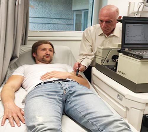
Abraham Katzir, PhD, a physics professor at Tel Aviv University, has developed a fiber-optic evanescent wave spectroscopy (FEWS) system based on middle infrared transmitting AgCIBr fibers and a Fourier-transform infrared spectrometer that determines the properties of the various skin lesions and identifies them based on their coloration within the infrared spectrum.

Tel Aviv University Physics Professor Abraham Katzir, PhD (above), demonstrates his new method of detecting cancerous skin lesion, which employs infrared sensors and optical fibers to determine the properties of various lesions on the skin and identify them based on their coloration within the infrared spectrum. (Photo copyright: Tel Aviv University.)
“We figured that with the help of devices that can identify these colors, healthy skin and each of the benign and malignant lesions would have different ‘colors,’ which would enable us to identify melanoma,” Katzir said in an Israel 21c news article.
The Israeli researchers published their study of 100 patients at a major Israeli hospital in Medical Physics, a journal of the American Association of Physicists in Medicine (AAPM).
“Melanoma is a life-threatening cancer, so it is very important to diagnose it early on, when it is still superficial,” Katzir told Israel 21c, adding that the new technology has the potential to cause “dramatic change” in the field of diagnosing and treating skin cancer, “and perhaps other types of cancer as well.”
As advancements in the non-invasive diagnosis of skin cancers continue, dermatopathologists—and in fact all anatomic and histopathology practices—should prepare for the financial impact this change may have on their clinical practices as demand for skin biopsies decreases.
—Andrea Downing Peck
Related Information
Novel Method Develop by Lithuanian Scientists Can Reach More than 90% Accuracy in Detecting Melanoma
Scientist Develops Instant Non-Invasive Cancer Detection Technology


