


Syringe-based technique is disposable and enables clinical laboratories to process small biopsies in about two hours instead of overnight and with significantly less waste
Histotechnologists and clinical laboratory managers know that the standard method of processing tissue biopsies takes a lot of time and chemical resources and isn’t always efficient. But what if there was a way to process biopsy tissue without the need for large processors that require a large batch of tissue to be economical?
That is what cytopathologist Paul Lee, MD, PhD, Assistant Professor of Clinical Pathology, University of Cincinnati (UC) College of Medicine, asked himself when he set out to design a rapid technique for processing small biopsies.
Lee was inspired to find a way to change the process while completing his residency at the University of Massachusetts, UC News reported.
“I noticed a specific issue with the procedure for fixing and examining tissue samples to look for signs of cancer and other diseases,” Lee told UC News. “And I had this idea.”
His goal was to reduce time to answer for a patient waiting to learn if he/she has cancer.
To achieve this feat, Lee developed a new technique that, according to UC News, “employs a disposable syringe and cuvette to do individual tissue tests, using small paraffin blocks and a combined embedding-fixing process for quick, accurate reads of small biopsies.”
Lee says his technique brings the potential of “immediate reads” closer to reality.
“If that process can take just two hours, not overnight, it becomes an inpatient procedure,” Lee told UC News. “Patients don’t have to go home … and return for a surgery consult, then for surgery itself.
“All that can be arranged in a day or two,” he added. “Patient care won’t be compromised or lost to follow-up.”
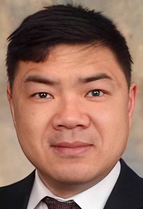
Paul Lee, MD, PhD (above), Assistant Professor of Clinical Pathology at University of Cincinnati College of Medicine, compares the development of his new small-biopsy tissue processing technique for histology laboratories and clinical laboratories to the philosophy behind the invention of the Keurig single-serving beverage machine. “Let’s say you’re making a cup of coffee. If you made a whole carafe and only needed one cup, that’d be wasteful—of both time and resources. Think of this as Keurig for specimen processing.” (Photo copyright: University of Cincinnati.)
Simplifying, Accelerating Rapid Tissue Processing
Lee describes the traditional method “coupling large tissue processors with traditional embedding techniques” as “slow and wasteful.” This, he told UC News, is still how tissue processing is done.
“It [uses] huge amounts of solvent, massive paraffin blocks,” he continued, “and [leaves] doctors waiting up to seven hours for results.”
The standard procedure uses “an enormous processor, gallons of solvent, and 300-500 dehydrated specimens embedded in blocks and then cut into slices for slides,” he added.
In addition to the “waste or expense,” the process “prevents physicians from making same-day diagnoses unless they’re willing to destroy precious tissue,” Lee noted.
Lee told UC News that his technique “preserves tissue [and] doesn’t compromise the sample, so we can do ancillary tests to revalidate results … and with the disposable cuvette there’s no chance of cross-contamination. Plus, it can be easily incorporated into existing infrastructure. [It] doesn’t have to upset processes or workflow.”
Lee’s method can also save resources and reduce wait times. “I get requests [from other researchers] all the time for various samples and I have to put a lot of them off for human pathology tests,” Lee said. “They can be their own processors and not wait for results from another lab. It’s quicker for them too and uses fewer resources.”
Other Advantages of Lee’s Method
Lee’s research team has successfully tested a prototype and they are currently awaiting a patent.
According to UC’s Office of Innovation, advantages of Lee’s new technique for small-biopsy tissue processing include:
- Rapid, convenient processing.
- Disposable specimen cuvette (no cross contamination).
- Antigenicity preservation.
- Less solvent usage (associated with less cost for solvent disposal).
- Can be easily incorporated into existing infrastructure.
- Very small footprint.
“Turn-around times for ‘rapid processing’ using current techniques typically range from four to seven hours, often preventing physicians from making same day diagnosis without destroying precious tissue,” the Office of Innovation noted in a statement. “This often results in delayed diagnosis, additional use of both patient and healthcare resources, and potentially poorer patient outcomes.
“Dr. Paul Lee has developed a novel tissue fixation and embedding system that combines the tissue fixation and embedding process creating a rapid processing block for biological specimens,” UC’s Office of Innovation continued. “The invention dramatically shortens processing and embedding time to approximately two hours while preserving the antigenicity and morphology of the specimen and thus allows for rapid reads of small biopsies in a timeframe that was not previously achievable.”
Lee’s work could streamline tissue processing in histology laboratories and increase efficiency without sacrificing accuracy. Anatomic pathologists and clinical laboratories would be wise to monitor this revolutionary new technology for further developments.
—Ashley Croce
Related Information: