New study shows how artificial neural networks are leading to improved microscopic capabilities and precision that could help researchers better understand causes of tumors
Biophysicists at the École Polytechnique Fédérale de Lausanne (EPFL) in Switzerland have developed a method for using artificial neural networks to automate control of clinical laboratory microscopes involved in a range of diagnostic testing.
Pathologists, microbiologists, and clinical laboratory scientists engaged in research into cellular activity will be interested to learn that advances in artificial intelligence (AI) are leading to new opportunities for future research studies to observe and capture—in real time—biological processes in living cells/samples.
“EPFL biophysicists have now found a way to automate microscope control for imaging biological events in detail while limiting stress on the sample, all with the help of artificial neural networks. Their technique works for bacterial cell division, and for mitochondrial division,” according to Phys.org.
Even better, the EPFL scientists are making the control framework available as an open-source software plug-in for the microscope control program Micro-Manager. This enables clinical laboratories to integrate artificial intelligence into existing microscope control software, Phys.org noted.
The researchers published their findings in the journal Nature Methods, titled, “Event-Driven Acquisition for Content-Enriched Microscopy.”

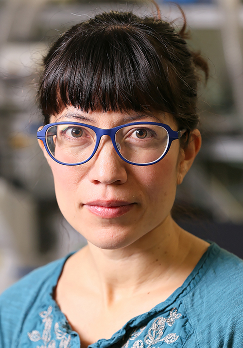
“An intelligent microscope is kind of like a self-driving car. It needs to process certain types of information, subtle patterns that it then responds to by changing its behavior,” Suliana Manley, PhD, biophysicist and a professor in the Laboratory of Experimental Biophysics at the École Polytechnique Fédérale de Lausanne, told Phys.org. Clinical laboratories and anatomic pathology groups may want to explore the capabilities of this intelligent microscope technology. (Photo copyright: EPFL.)
EPFL Study Breakdown
Suliana Manley, PhD, biophysicist, professor, and head of the Laboratory of Experimental Biophysics at the EPFL, is the principal investigator of the “Intelligent microscope” study. She has spent her career focusing on the development of high-resolution optical instruments and the organization and dynamics of proteins.
Manley and her team learned how to detect mitochondrial divisions in bacteria such as Caulobacter Crescentus. These divisions can occur once every few minutes, but last only seconds. Their infrequency and ability to occur anywhere within the mitochondrial network makes them difficult to spot and photograph, Physics World noted.
To overcome this barrier, the EPFL team trained the neural network to look for mitochondrial constrictions—the shape change in mitochondria that leads to division—and combined that data with observations of the protein DRP1, the presence of which is required for spontaneous divisions to occur, Phys.org reported.
“A common goal of fluorescence microscopy is to collect data on specific biological events. Yet, the event-specific content that can be collected from a sample is limited, especially for rare or stochastic processes,” wrote the EPFL scientists in their Nature Methods paper. “We developed an event-driven acquisition framework, in which neural-network-based recognition of specific biological events triggers real-time control in an instant structured illumination microscope … we capture mitochondrial and bacterial divisions at imaging rates that match their dynamic timescales, while extending overall imaging durations. Because event-driven acquisition allows the microscope to respond specifically to complex biological events, it acquires data enriched in relevant content.”
Manley’s intelligent fluorescent microscope responds more rapidly than human control can achieve. When the microscope senses constrictions and DRP1 protein levels are high, it switches into high-speed image capture mode and gathers multiple images with minute detail. Conversely, when both constrictions and protein are low, the microscope goes into low-speed imaging mode, which preserves the integrity of the sample by avoiding excessive exposure to light, Phys.org noted.
Automating microscope control limits the stress placed on samples, increasing the likelihood of more accurate results. It also allows microscope control for imaging biological events in detail, as it eliminates human error that naturally occurs from trying to keep up with events in real time, Phys.org reported.
“Using a neural network, we can detect much more subtle events and use them to drive changes in acquisition speed,” Manley told Phys.org. “The potential of intelligent microscopy includes measuring what standard acquisitions would miss. We capture more events, measure smaller constrictions, and can follow each division in greater detail.”
What’s Next for EPFL?
Manley and her EPFL team plan to continue working with neural networks to detect different events and bring about different hardware responses.
“For example, we envision harnessing optogenetic perturbations to modulate transcription at key moments in cell differentiation,” she told Physics World. “We also think of using event detection as a means of data compression, selecting for storage or analysis the pieces of data that are most relevant to a given study.”
As research continues in bacterial cell division, there will be a point where the technology enables researchers to observe cell activity and what conditions cause abnormal (tumor) cells to be created. That would be the first step to then investigating ways to stop the cellular process that creates abnormal cells.
It should not surprise pathologists and clinical laboratory managers that researchers and technology developers are exploring ways to “turbocharge” classic light microscopy. Advances in image analysis, combined with machine learning algorithms, are making it possible to tease new insights from the images viewed with a standard microscope.
—Kristin Althea O’Connor
Related Information:
Intelligent Microscope Uses AI to Capture Rare Biological Events
Intelligent Microscopes for Detecting Rare Biological Events


