Genetic data captured by this new technology could lead to a new understanding of how different types of cells exchange information and would be a boon to anatomic pathology research worldwide
What if it were possible to map the interior of cells and view their genetic sequences using chemicals instead of light? Might that spark an entirely new way of studying human physiology? That’s what researchers at the Massachusetts Institute of Technology (MIT) believe. They have developed a new approach to visualizing cells and tissues that could enable the development of entirely new anatomic pathology tests that target a broad range of cancers and diseases.
Scientists at MIT’s Broad Institute and McGovern Institute for Brain Research developed this new technique, which they call DNA Microscopy. They published their findings in Cell, titled, “DNA Microscopy: Optics-free Spatio-genetic Imaging by a Stand-Alone Chemical Reaction.”
Joshua Weinstein, PhD, a postdoctoral associate at the Broad Institute and first author of the study, said in a news release that DNA microscopy “is an entirely new way of visualizing cells that captures both spatial and genetic information simultaneously from a single specimen. It will allow us to see how genetically unique cells—those comprising the immune system, cancer, or the gut for instance—interact with one another and give rise to complex multicellular life.”
The news release goes on to state that the new technology “shows how biomolecules such as DNA and RNA are organized in cells and tissues, revealing spatial and molecular information that is not easily accessible through other microscopy methods. DNA microscopy also does not require specialized equipment, enabling large numbers of samples to be processed simultaneously.”

New Way to Visualize Cells
The MIT researchers saw an opportunity for DNA microscopy to find genomic-level cell information. They claim that DNA microscopy images cells from the inside and enables the capture of more data than with traditional light microscopy. Their new technique is a chemical-encoded approach to mapping cells that derives critical genetic insights from the organization of the DNA and RNA in cells and tissue.
And that type of genetic information could lead to new precision medicine treatments for chronic disease. New Atlas notes that “ Speeding the development of immunotherapy treatments by identifying the immune cells best suited to target a particular cancer cell is but one of the many potential application for DNA microscopy.”
In their published study, the scientists note that “Despite enormous progress in molecular profiling of cellular constituents, spatially mapping [cells] remains a disjointed and specialized machinery-intensive process, relying on either light microscopy or direct physical registration. Here, we demonstrate DNA microscopy, a distinct imaging modality for scalable, optics-free mapping of relative biomolecule positions.”
How DNA Microscopy Works
The New York Times (NYT) notes that the advantage of DNA microscopy is “that it combines spatial details with scientists’ growing interest in—and ability to measure—precise genomic sequences, much as Google Street View integrates restaurant names and reviews into outlines of city blocks.”
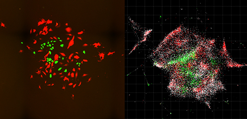
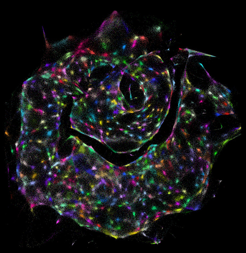
And Singularity Hub notes that “ DNA microscopy, uses only a pipette and some liquid reagents. Rather than monitoring photons, here the team relies on ‘bar codes’ that chemically tag onto biomolecules. Like cell phone towers, the tags amplify, broadcasting their signals outward. An algorithm can then piece together the captured location data and transform those GPS-like digits into rainbow-colored photos. The results are absolutely breathtaking. Cells shine like stars in a nebula, each pseudo-colored according to their genomic profiles.”
“We’ve used DNA in a way that’s mathematically similar to photons in light microscopy,” Weinstein said in the Broad Institute news release. “This allows us to visualize biology as cells see it and not as the human eye does.”
In their study, researchers used DNA microscopy to tag RNA molecules and map locations of individual human cancer cells. Their method is “surprisingly simple” New Atlas reported. Here’s how it’s done, according to the MIT news release:
- Small synthetic DNA tags (dubbed “barcodes” by the MIT team) are added to biological samples;
- The “tags” latch onto molecules of genetic material in the cells;
- The tags are then replicated through a chemical reaction;
- The tags combine and create more unique DNA labels;
- The scientists use a DNA sequencer to decode and reconstruct the biomolecules;
- A computer algorithm decodes the data and converts it to images displaying the biomolecules’ positions within the cells.

“The first time I saw a DNA microscopy image, it blew me away,” said Aviv Regev, PhD, a biologist at the Broad Institute, a Howard Hughes Medical Institute (HHMI) Investigator, and co-author of the MIT study, in an HHMI news release. “It’s an entirely new category of microscopy. It’s not just a technique; it’s a way of doing things that we haven’t ever considered doing before.”
Precision Medicine Potential
“Every cell has a unique make-up of DNA letters or genotype. By capturing information directly from the molecules being studied, DNA microscopy opens up a new way of connecting genotype to phenotype,” said Feng Zhang, PhD, MIT Neuroscience Professor,
Core Institute Member of the Broad Institute, and Investigator at the McGovern Institute for Brain Research at MIT, in the HHMI news release.
In other words, DNA microscopy could someday have applications in precision medicine. The MIT researchers, according to Stat, plan to expand the technology further to include immune cells that target cancer.
The Broad Institute has applied for a patent on DNA microscopy. Clinical laboratory and anatomic pathology group leaders seeking novel resources for diagnosis and treatment of cancer may want to follow the MIT scientists’ progress.
—Donna Marie Pocius
Related Information:
A Chemical Approach to Imaging Cells from the Inside
DNA Microscope Sees “Through the Eyes of the Cell”
DNA Microscopy Offers Entirely New Way to Image Cells
DNA Microscopy: Optics-free Spatio-Genetic Imaging by a Stand-Alone Chemical Reaction
This New Radical DNA Microscope Reimagines the Cellular World


