Nov 28, 2018 | Digital Pathology, Instruments & Equipment, Laboratory Instruments & Laboratory Equipment, Laboratory Management and Operations, Laboratory News, Laboratory Operations, Laboratory Pathology, Laboratory Testing
Clinical laboratory leaders aiming for patient-centered care and precision medicine outcomes need to acknowledge that patients do not want to be in hospitals or travel to physician offices and patient care centers for blood tests. It can be inconvenient, sometimes costly, and often painful.
That’s why disease management methods such as remote patient monitoring are appealing to many people. It’s a big market estimated to reach $1 billion by 2020, according to a Transparency Market Research Report. The study also associated popularity of devices such as heart rate and respiratory rate monitors with economic pressures of unnecessary hospital readmissions.
But can remote patient monitoring be used for more than to check heart rates, monitor blood glucose, and track activity levels? Could such technology be effectively leveraged by medical laboratories for remote blood sampling?
Microsampling versus Dried Blood Collecting
Remote patient monitoring must be able to address a large number of diseases and chronic health conditions for it to continue to expand and gain acceptance as a viable way to care for patients in different settings outside of hospitals. However, as most clinical pathologists and laboratory scientists know, clinical laboratory testing has an essential role in patient monitoring. Thus, there is the need for a way to collect blood and other relevant samples from patients in these remote settings.
One promising approach is the development of new microsampling technology that can overcome past obstacles of dried blood collection. Furthermore, microsampling-enabled devices can make it possible for medical laboratories to reach out to the homebound to secure accurate and volumetrically appropriate samples in a cost-effective manner.
“One well-established fact in today’s healthcare system is that an ever-greater proportion of patients want clinical care that is less invasive and less intrusive,” noted Robert Michel, Editor-in-Chief of Dark Daily and The Dark Report. “Patients want to take more control over their treatment and be more effective at maintaining the stability of their chronic conditions, and often are happier than those who need to travel to have chronic conditions monitored. To meet this need there has been significant innovation, particularly in the area of remote blood sampling using microsampling technology.”
For decades, medical laboratories have tried various methods for acquiring and transporting blood samples from remote locations. One such non-invasive alternative to venipuncture is called dried blood spot (DBS) collecting. It involves placing a fingerprick of blood on filter paper and allowing it to dry prior to transport to the lab.
But DBS collected bio samples often do not contain enough hematocrit (volume percentage of red blood cells) for laboratories and clinical pathologists to provide accurate reports and interpretations. Reported reasons DBS cards have not penetrated a wide market include:
- Hematocrit bias or effect;
- Costly card punching and automation equipment; and,
- Possible disruption to existing lab workflows.
Microsampling Technology Enables Collection of Appropriate Samples
Microsampling has to have the capability to enable labs to deliver quality results from reliable blood samples. This remote sampling technology makes it possible for phlebotomists to offer a comfortable collection alternative for homebound patients and rural residents. It also can be useful for physicians stationed in remote areas. Patients themselves can even collect their own blood samples.
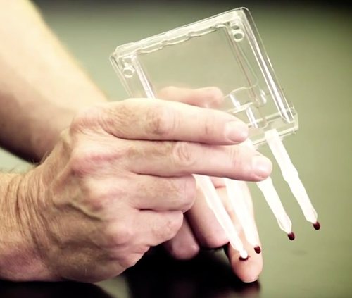
Volumetric Absorptive Microsampling (VAMS) technology enables accurate samples of blood or other fluids from amounts as small as 10, 20, or 30 microliters, according to Neoteryx, LLC, of Torrance, Calif., the developer of VAMS. The technology is integrated into the company’s Mitra microsampler blood collection devices (shown above) in formats for patient use and for medical laboratory microsample accessioning and extraction. Click here to watch a video on the Mitra Microsampler Specimen Collection Device. (Photo copyright: Neoteryx.)
One company developing these types of products is Neoteryx, LLC, of Torrance, Calif. It develops, manufactures, and distributes microsampling products. Patients with the company’s Mitra device use a lancet to puncture their skin and draw a small amount of blood, collect it on the device’s absorptive tip, and then mail the samples to a blood lab for testing (Neoteryx does not perform testing).
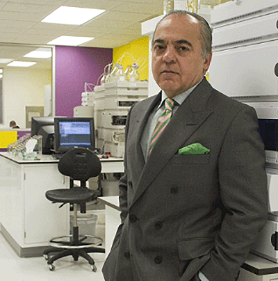
“Technologies such VAMS are driving [precision medicine] in an extremely cost-effective manner, while only requiring minimal patient effort. Patients are taking a more active role in their healthcare journeys, and at-home sampling is supporting this shift,” stated Fasha Mahjoor, Chief Executive Officer, Neoteryx, in a blog post. (Photo copyright: Neoteryx.)
Patient satisfaction survey data collected by Neoteryx suggest patients are comfortable with their role in blood collection:
- 70% are comfortable or very comfortable with the process;
- 86% say it is easy or very easy to use the Mitra device;
- 92% report it is easy to capture blood on the device’s tip;
- 55% of Mitra device users are likely or very likely to choose microsampling over traditional venipuncture; and,
- 93% noted they are likely or very likely to choose the device for child care.
A list of published studies describes certain advantages of VAMS technology that have implications for medical laboratories and clinical pathologists:
- Microsampling has benefits and implications for therapeutic drug monitoring, infectious disease research, and remote specimen collection;
- Dried blood microsamples from fingerstick can generate reliable data “correlating” to traditional blood collection processes;
- Bioanalytical data collected with the Mitra device are accurate and dependable; and,
- In a study for a panel of anti-epileptic drugs, VAMS led to optimized extraction efficiency above 86%, which means there was no hematocrit bias.
Learn More by Requesting the Dark Daily Microsampling White Paper
To help medical laboratories and clinical pathologists learn more about microsampling and VAMS devices, Dark Daily and The Dark Report have produced a white paper titled “How to Create a Patient-Centered Lab with Breakthrough Blood Collection Technology: Microsampling Takes Blood Collection Out of the Clinic.” The paper includes sections addressing these topics:
- Rise of patient-centered care and remote patient monitoring;
- Dried blood collection over the years and the hematocrit effect;
- A look at microsampling and how it takes blood collection out of the clinic;
- How Volumetric Absorptive Microsampling (VAMS) technology works;
- Patient satisfaction data;
- Research about microsampling including extensive graphics;
- Launching new VAMS technology; and,
- Frequently asked questions.
Innovative medical laboratory leaders who want to increase their understanding of how microsampling technology and remote patient monitoring relates to the goal of becoming a patient-centered lab are encouraged to request a copy of the white paper. It can be downloaded at no cost by clicking here, or placing https://www.darkdaily.com/how-to-create-a-patient-centered-lab-with-breakthrough-blood-collection-technology-9-2018/ into your browser.
—Donna Marie Pocius
Related Information:
Remote Patient Monitoring Devices Market
Neoteryx, LLC, and Cedars Sinai Partner to Investigate at Home Blood Sampling Possibilities for Patients with Inflammatory Bowel Disease
Creating a Patient-Centered Lab with Breakthrough Blood Collection Technology Using New Microsampling Methods Provides Reliable, Economic Collection, Shipping and Storage Solutions
How to Create a Patient-Centered Lab with Breakthrough Blood Collection Technology: Microscopy Takes Blood Collection Out of the Clinic
Nov 16, 2018 | Digital Pathology, Instruments & Equipment, Laboratory Instruments & Laboratory Equipment, Laboratory Management and Operations, Laboratory News, Laboratory Operations, Laboratory Pathology, Laboratory Testing
New study conducted by an international team of researchers suggests that artificial intelligence (AI) may be better than highly-trained humans at detecting certain skin cancers
Artificial intelligence (AI) has been working its way into health technology for several years and, so far, AI tools have been a boon to physicians and health networks. Until now, though, the general view was that it was a supplemental tool for diagnosticians, not a replacement for them. But what if the AI was better at detecting disease than humans, including anatomic pathologists?
Researchers in the Department of Dermatology at Heidelberg University in Germany have concluded that AI can be more accurate at identifying certain cancers. The challenge they designed for their study involved skin biopsies and dermatologists.
They pitted a deep-learning convolutional neural network (CNN) against 58 dermatologists from 17 countries to determine which was more accurate at detecting malignant melanomas—humans or AI. A CNN is an artificial network based on the biological processes that occur when neurons in the brain are connected to each other and respond to what the eye sees.
The CNN won.
“For the first time we compared a CNN’s diagnostic performance with a large international group of 58 dermatologists, including 30 experts. Most dermatologists were outperformed by the CNN. Irrespective of any physicians’ experience, they may benefit from assistance by a CNN’s image classification,” the report noted.
The researchers published their report in the Annals of Oncology, a peer-reviewed medical journal published by Oxford University Press that is the official journal of the European Society for Medical Oncology.

“I expected only a performance on an even level with the physicians. The outperformance even of the average experienced and trained dermatologists was a major surprise,” Holger Haenssle, PhD, Professor of Dermatology at Heidelberg University and one of the authors of the study, told Healthline. Anatomic pathologists will want to follow the further development of this research and its associated diagnostic technologies. (Photo copyright: University of Heidelberg.)
Does AI Tech Have Superior Visual Acuity Compared to Human Eyes?
The dermatologists who participated in the study had varying degrees of experience in dermoscopy, also known as dermatoscopy. Thirty of the doctors had more than five-year’s experience and were considered to be expert level. Eleven of the dermatologists were considered “skilled” with two- to five-year’s experience. The remaining 17 doctors were termed beginners with less than two-year’s experience.
To perform the study, the researchers first compiled a set of 100 dermoscopic images that showed melanomas and benign moles called Nevi. Dermoscopes (or dermatoscopes) create images using a magnifying glass and light source pressed against the skin. The resulting magnified, high-resolution images allow for easier, more accurate diagnoses than inspection with the naked eye.
During the first stage of the research, the dermatologists were asked to diagnose whether a lesion was melanoma or benign by looking at the images with their naked eyes. They also were asked to render their opinions for any needed action, such as surgery and follow-up care based on their diagnoses.
After this part of the study, the dermatologists on average identified 86.6% of the melanomas and 71.3% of the benign moles. More experienced doctors identified the melanomas at 89%, which was slightly higher than the average of the group.
The researchers also showed 300 images of malignant and benign skin lesions to the CNN. The AI accurately identified 95% of the melanomas by analyzing the images.
“The CNN missed fewer melanomas, meaning it had a higher sensitivity than the dermatologists, and it misdiagnosed fewer benign moles as malignant melanoma, which means it had a higher specificity. This would result in less unnecessary surgery,” Haenssle told CBS News.
In a later part of the research, the dermatologists were shown the images a second time and provided clinical information about the patients, including age, gender, and location of the lesion. They were again instructed to make diagnoses and projected care decisions. With the additional information, the doctors’ average detection of melanomas increased to 88.9% and their recognition of benign moles increased to 75.7%. Still below the results of the CNN.
These findings suggest that the visual pattern recognition of AI technology could be a meaningful tool to help physicians and researchers diagnose certain cancers.
“In the future, I think AI will be integrated into practice as a diagnostic aide, particularly in primary care, to support the decision to excise a lesion, refer, or otherwise to reassure that it is benign,” Victoria Mar, PhD, an Adjunct Senior Lecturer in the Department of Public Health and Preventative Medicine at Australia’s Monash University, told Healthline.
“There is the potential for AI technology to be integrated with 2D or 3D skin imaging systems, which means that the majority of benign lesions would be already filtered by the machine, so that we can spend more time concentrating on the difficult or more concerning lesions,” she said. “To me, this means a more productive interaction with the patient, where we can focus on appropriate management and provide more streamlined care.”
AI Performs Well in Other Studies Involving Skin Biopsies
This study is not the only research that suggests entities besides humans may be utilized in diagnosing some cancers from images. Last year, computer scientists at Stanford University performed similar research and found comparable results. For that study, the researchers created and trained an algorithm to visually diagnose potential skin cancers by looking at a database of skin images. They then showed photos of skin lesions to 21 dermatologists and asked for their diagnoses based on the images. They found the accuracy of their AI matched the performance of the doctors when diagnosing skin cancer from viewed images.
And in 2017, Dark Daily reported on three genomic companies developing AI/facial recognition software that could help anatomic pathologists diagnose rare genetic disorders. (See, “Genomic Companies Collaborate to Develop Facial Analysis Technology Pathologists Might Eventually Use to Diagnose Rare Genetic Disorders,” August 7, 2017.)
While many dermatologists read patient biopsies on their own, they also refer high volumes of skin biopsies to anatomic pathologists. A technology that can accurately diagnose skin cancers could potentially impact the workload received by clinical laboratories and anatomic pathology groups.
—JP Schlingman
Related Information:
Dermatologists Hate Him! Meet the Skin-cancer Detecting Robot
Man Against Machine: Diagnostic Performance of a Deep Learning Convolutional Neural Network for Dermoscopic Melanoma Recognition in Comparison to 58 Dermatologists
AI Better than Dermatologists at Detecting Skin Cancer, Study Finds
AI May Be Better at Detecting Skin Cancer than Your Derm
Deep Learning Algorithm Does as Well as Dermatologists in Identifying Skin Cancer
Genomic Companies Collaborate to Develop Facial Analysis Technology Pathologists Might Eventually Use to Diagnose Rare Genetic Disorders
Nov 5, 2018 | Digital Pathology, Instruments & Equipment, Laboratory Instruments & Laboratory Equipment, Laboratory Management and Operations, Laboratory News, Laboratory Operations, Laboratory Pathology, Laboratory Testing
Computer-assisted analysis using Google’s LYNA algorithm shows significant gains in speed required to analyze stained lymph node slides and sensitivity of micrometastases detection in two recent studies
Anatomic pathologists understand the complexities of reviewing slides and samples for signs of cancer’s spread. Two studies involving a new artificial intelligence (AI) algorithm from Google (NASDAQ:GOOGL) claim their “deep learning” LYmph Node Assistant (LYNA) provides increases to both the speed at which pathologists can analyze slides and improved accuracy in detecting metastatic breast cancer within the slide samples used for the studies.
Google’s first study was published in the Archives of Pathology and Laboratory Medicine and investigated the accuracy of the algorithm using digital pathology slides. Google’s second study, published in The American Journal of Surgical Pathology, looked at how pathologists might harness the algorithm to improve workflows and use the tool in a clinical setting.
Medical laboratories and other diagnostics providers are already familiar with the improvement potential of automation and other technology-based approaches to diagnosis and analysis. Google’s LYNA is an example of how AI and machine learning improvements can serve as a supplement to—not a replacement for—the skills of experts at pathology groups and clinical laboratories.
Early research done by Google indicates that integrating LYNA into existing workflows could allow pathologists to spend less time analyzing slides for minute details. Instead, they could focus on other more challenging tasks while the AI analyzes gigapixels worth of slide data to highlight regions of concern in slides and samples for deeper manual inspection.
LYNA Achieves 99% Accuracy in Study of Metastatic Breast Cancer Detection
According to the research cited in a Google AI Blog post, roughly 25% of metastatic lymph node staging classifications would change if subjected to a second pathologic review. They further note that when faced with time constraints, detection sensitivity for small metastases on individual slides can be as low as 38%.
In findings published in Archives of Pathology and Laboratory Medicine, Google researchers analyzed whole slide images from hematoxylin-eosin-stained lymph nodes for 399 patients sourced from the Camelyon16 challenge dataset. Of those slides, researchers used 270 to train LYNA and the remaining 129 for analysis. They then compared the LYNA findings to those of an independent lab using a different scanner.
“LYNA achieved a slide-level area under the receiver operating characteristic (AUC) of 99% and a tumor-level sensitivity of 91% at one false positive per patient on the Camelyon16 evaluation dataset,” the researchers stated. “We also identified [two] ‘normal’ slides that contained micrometastases.”
Google’s algorithm later received an AUC of 99.6% on a secondary dataset.
“Artificial intelligence algorithms can exhaustively evaluate every tissue patch on a slide, achieving higher tumor-level sensitivity than, and comparable slide-level performance to, pathologists,” the researchers continued. “These techniques may improve the pathologist’s productivity and reduce the number of false negatives associated with morphologic detection of tumor cells.”
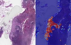
Left: sample view of a slide containing lymph nodes, with multiple artifacts: the dark zone on the left is an air bubble, the white streaks are cutting artifacts, the red hue across some regions are hemorrhagic (containing blood), the tissue is necrotic (decaying), and the processing quality was poor. Right: LYNA identifies the tumor region in the center (red), and correctly classifies the surrounding artifact-laden regions as non-tumor (blue). (Image and caption copyright: Google AI Blog.)
Faster Analysis through Software Assistance
Rapid diagnosis helps improve cancer outcomes. Yet, manually reviewing and analyzing complex digital slides is time-consuming. Time constraints might also lead to false negatives due to micrometastases or small suspicious regions that slip by pathologists undetected.
The Google research team of the study published in The American Journal of Surgical Pathology sought to gauge the impact LYNA might have on the histopathologic review of lymph nodes for trained pathologists. In their multi-reader multi-case study, researchers analyzed differences in both sensitivity of detecting micrometastases and the average review time per image using both computer-aided detection and unassisted detection for six pathologists across 70 slides.
Using the LYNA algorithm to identify and outline regions likely to contain tumors, the researchers found that sensitivity increased from 83% to 91%. The time to review slides also saw a significant reduction from 116 seconds in the unassisted mode to 61 seconds in the assisted mode—a time savings of roughly 47%.
“Although some pathologists in the unassisted mode were less sensitive than LYNA,” the researchers stated, “all pathologists performed better than the algorithm alone in regard to both sensitivity and specificity when reviewing images with assistance.”
The Future of Digital Pathology using LYNA
While the two studies show positive results, both studies also reveal shortcomings. Google highlighted both limited dataset sizes and simulated diagnostic workflows as potential concerns and areas on which to focus future studies.
Still, Google’s researchers believe that algorithms such as LYNA will help to power the future of diagnostics as healthcare in the digital era continues to mature. “We remain optimistic,” state the authors of the Google AI Blog post, “that carefully validated deep learning technologies and well-designed clinical tools can help improve both the accuracy and availability of pathologic diagnosis around the world.”
While other industries see risk in the growth of AI, both studies performed by researchers at Google show how computer-assisted workflows and machine learning could accentuate and bolster the skills of trained diagnosticians, such as anatomic pathologists and clinical laboratory technicians. By working to compensate for weak points in both human skill and computer reasoning, the outcome could be greater than either AI or humans can achieve separately.
—Jon Stone
Related Information:
Google Creates AI to Detect When Breast Cancer Spreads
Google Deep Learning Tool 99% Accurate at Breast Cancer Detection
Google’s AI Software Seeks to Detect Advanced Breast Cancer Better Than We Have Before
Google’s AI Is Better at Spotting Advanced Breast Cancer than Pathologists
Google AI Claims 99% Accuracy in Metastatic Breast Cancer Detection
Applying Deep Learning to Metastatic Breast Cancer Detection
Assisting Pathologists in Detecting Cancer with Deep Learning
Diagnostic Assessment of Deep Learning Algorithms for Detection of Lymph Node Metastases in Women with Breast Cancer
Nov 2, 2018 | Coding, Billing, and Collections, Compliance, Legal, and Malpractice, Digital Pathology, Instruments & Equipment, Laboratory Instruments & Laboratory Equipment, Laboratory Management and Operations, Laboratory News, Laboratory Operations, Laboratory Pathology, Laboratory Testing, Management & Operations
Patient privacy, ethics of monetizing not-for-profit data, and questions surrounding industry conflicts appear after the public announcement of an arrangement to grant exclusive access to academic pathology slides and samples
Clinical laboratories and anatomic pathology groups already serve as gatekeepers for a range of medical data used in patient treatments. Glass slides, paraffin-embedded tissue specimens, pathology reports, and autopsy records hold immense value to researchers. The challenge has been how pathologists (and others) in a not-for-profit academic center could set themselves up to potentially profit from their exclusive access to this archived pathology material.
Now, a recent partnership between Memorial Sloan Kettering Cancer Center (MSK) and Paige.AI (a developer of artificial intelligence for pathology) shows how academic pathology laboratories might accomplish this goal and serve a similar gatekeeper role in research and development using the decades of cases in their archives.
The arrangement, however, is not without controversy.
New York Times, ProPublica Report
Following an investigative report from the New York Times (NYT) and ProPublica, pathologists and board members at MSK are under fire from doctors and scientists there who have concerns surrounding ethics, exclusivity, and profiting from data generated by physicians and but owned by MSK.
“Hospital pathologists have strongly objected to the Paige.AI deal, saying it is unfair that the founders received equity stakes in a company that relies on the pathologists’ expertise and work amassed over 60 years. They also questioned the use of patients’ data—even if it is anonymous—without their knowledge in a profit-driven venture,” the NYT article states.
Prominent members of MSK are facing scrutiny from the media and peers—with some relinquishing stakes in Paige.AI—as part of the backlash of the report. This is an example of the perils and PR concerns lab stakeholders might face concerning the safety of data sharing and profits made by medical laboratories and other diagnostics providers using patient data.
Controversy Surrounds Formation of Paige.AI/MSK Partnership
In February 2018, Paige.AI announced closing the deal on a $25-million round of Series A funding, and in gaining exclusive access to 25-million pathology slides and computational pathology intellectual property held by the Department of Pathology at Memorial Sloan Kettering. Coverage by TechCrunch noted that while MSK received an equity stake as part of the licensing agreement, they were not a cash investor.
Creation of the company involved three hospital insiders and three additional board members with the hospital itself established as part owner, according to STAT.
Unnamed officials told the NYT that board members at MSK only invested in Paige.AI after earlier efforts to generate outside interest and investors were unsuccessful. NYT’s coverage also notes experts in non-profit law and corporate governance have raised questions as to compliance with federal and state laws that govern nonprofits in light of the Paige.AI deal.
Growing Privacy Fallout and Potential Pitfalls for Medical Labs
The original September 2018 NYT coverage noted that Klimstra intends to divest his ownership stake in Paige.AI. Later coverage by NYT in October, notes that Democrat Representative Debbie Dingell of Michigan submitted a letter questioning details about patient privacy related to Paige.AI’s access to MSK’s academic pathology resources.
Privacy continues to be a focus for both media and regulatory scrutiny as patient data continues to fill electronic health record (EHR) systems as well as research and commercial databases. Dark Daily recently covered how University of Melbourne researchers demonstrated how easily malicious parties might reidentify deidentified data. (See “Researchers Easily Reidentify Deidentified Patient Records with 95% Accuracy; Privacy Protection of Patient Test Records a Concern for Clinical Laboratories”, October 10, 2018.)
According to the NYT, MSK also issued a memo to employees announcing new restrictions on interactions with for-profit companies with a moratorium on board members investing in or holding board positions in startups created within MSK. The nonprofit further noted it is considering barring hospital executives from receiving compensation for their work on outside boards.
However, MSK told the NYT this only applies to new deals and will not affect the exclusive deal between Paige.AI and MSK.
“We have determined,” MSK wrote, “that when profits emerge through the monetization of our research, financial payments to MSK-designated board members should be used for the benefit of the institution.”
There are no current official legal filings regarding actions against the partnership. Despite this, the arrangement—and the subsequent fallout after the public announcement of the arrangement—serve as an example of pitfalls medical laboratories and other medical service centers considering similar arrangements might face in terms of public relations and employee scrutiny.
Risk versus Reward of Monetizing Pathology Data
While the Paige.AI situation is only one of multiple concerns now facing healthcare teams and board members at MSK, the events are an example of risks pathologists take when playing a role in a commercial enterprise outside their own operations or departments.
In doing so, the pathologists investing in and shaping the deal with Paige.AI brought criticism from reputable sources and negative exposure in major media outlets for their enterprise, themselves, and MSK as a whole. The lesson from this episode is that pathologists should tread carefully when entertaining offers to access the patient materials and data archived by their respective anatomic pathology and clinical laboratory organizations.
—Jon Stone
Related Information:
Sloan Kettering’s Cozy Deal with Start-Up Ignites a New Uproar
Paige.AI Nabs $25M, Inks IP Deal with Sloan Kettering to Bring Machine Learning to Cancer Pathology
Sloan Kettering Executive Turns Over Windfall Stake in Biotech Start-Up
Cancer Center’s Board Chairman Faults Top Doctor over ‘Crossed Lines’
Memorial Sloan Kettering, You’ve Betrayed My Trust
LVHN Patient Data Not Shared with For-Profit Company in Sloan Kettering Trials
Researchers Easily Reidentify Deidentified Patient Records with 95% Accuracy; Privacy Protection of Patient Test Records a Concern for Clinical Laboratories
Oct 26, 2018 | Digital Pathology, Instruments & Equipment, Laboratory Instruments & Laboratory Equipment, Laboratory Management and Operations, Laboratory News, Laboratory Operations, Laboratory Pathology, Laboratory Testing
Future EHRs will focus on efficiency, machine learning, and cloud services—improving how physicians and medical laboratories interact with the systems to support precision medicine and streamlined workflows
When the next generation of electronic health record (EHR) systems reaches the market, they will have advanced features that include cloud-based services and the ability to collect data from and communicate with patients using mobile devices. These new developments will provide clinical laboratories and anatomic pathology groups with new opportunities to create value with their lab testing services.
Proposed Improvements and Key Trends
Experts with EHR developers Epic Systems, Allscripts, Accenture, and drchrono spoke recently with Healthcare IT News about future platform initiatives and trends they feel will shape their next generation of EHR offerings.
They include:
- Automation analytics and human-centered designs for increased efficiency and to help reduce physician burnout;
- Improved feature parity across mobile and computer EHR interfaces to provide patients, physicians, and medical laboratories with access to information across a range of technologies and locations;
- Integration of machine learning and predictive modeling to improve analytics and allow for better implementation of genomics-informed medicine and population health features; and
- A shift toward cloud-hosted EHR solutions with support for application programming interfaces (APIs) designed for specific healthcare facilities that reduce IT overhead and make EHR systems accessible to smaller practices and facilities.
Should these proposals move forward, future generations of EHR platforms could transform from simple data storage/retrieval systems into critical tools physicians and medical laboratories use to facilitate communications and support decision-making in real time.
And, cloud-based EHRs with access to clinical labs’ APIs could enable those laboratories to communicate with and receive data from EHR systems with greater efficiency. This would eliminate yet another bottleneck in the decision-making process, and help laboratories increase volumes and margins through reduced documentation and data management overhead.
Cloud-based EHRs and Potential Pitfalls
Cloud-based EHRs rely on cloud computing, where IT resources are shared among multiple entities over the Internet. Such EHRs are highly scalable and allow end users to save money by hiring third-party IT services, rather than maintaining expensive IT staff.
Kipp Webb, MD, provider practice lead and Chief Clinical Innovation Officer at Accenture told Healthcare IT News that several EHR vendors are only a few years out on releasing cloud-based inpatient/outpatient EHR systems capable of meeting the needs of full-service medical centers.
While such a system would mean existing health networks would not need private infrastructure and dedicate IT teams to manage EHR system operations, a major shift in how next-gen systems are deployed and maintained could lead to potential interoperability and data transmission concerns. At least in the short term.
Yet, the transition also could lead to improved flexibility and connectivity between health networks and data providers—such as clinical laboratories and pathologist groups. This would be achieved through application programming interfaces (APIs) that enable computer systems to talk to each other and exchange data much more efficiently.
“Perhaps one of the biggest ways having a fully cloud-based EHR will change the way we as an industry operate will be enabled API access.” Daniel Kivatinos, COO and founder of drchrono, told Healthcare IT News. “You will be able to add other partners into the mix that just weren’t available before when you have a local EHR install only.”

Paul Black, CEO of Allscripts, believes these changes will likely require more than upgrading existing software or hardware. “The industry needs an entirely new approach to the EHR,” he told Healthcare IT News. “We’re seeing a huge need for the EHR to be mobile, cloud-based, and comprehensive to streamline workflow and get smarter with every use.” (Photo copyright: Allscripts.)
Reducing Physician Burnout through Human-Centered Design
As Dark Daily reported last year, EHRs have been identified as contributing to physician burnout, increased dissatisfaction, and decreased face-to-face interactions with patients.
Combined with the increased automation, Carl Dvorak, President of Epic Systems, notes next-gen EHR changes hold the potential to streamline the communication of orders, laboratory testing data, and information relevant to patient care. They could help physicians reach treatment decisions faster and provide laboratories with more insight, so they can suggest appropriate testing pathways for each episode of care.
“[Automation analytics] holds the key to unlocking some of the secrets to physician well-being,” Dvorak told Healthcare IT News. “For example, we can avoid work being unnecessarily diverted to physicians when it could be better managed by others.”
Black echoes similar benefits, saying, “We believe using human-centered design will transform the way physicians experience and interact with technology, as well as improve provider wellness.”
Some might question the success of the first wave of EHR systems. Though primarily built to address healthcare reform requirements, these systems provided critical feedback and data to EHR developers focused not on simply fulfilling regulatory requirements, but on meeting the needs of patients and care providers as well.
If these next-generations systems can help improve the quality of data recording, storage, and transmission, while also reducing physician burnout, they will have come a long way from the early EHRs. For medical laboratory professionals, these changes will likely impact how orders are received and lab results are reported back to doctors in the future. Thus, it’s worth monitoring these developments.
—Jon Stone
Related Information:
Next-Gen EHRs: Epic, Allscripts and Others Reveal Future of Electronic Health Records
Next-Gen IT Infrastructure: A Nervous System Backed by Analytics and Context
EHR Systems Continue to Cause Burnout, Physician Dissatisfaction, and Decreased Face-to-Face Patient Care
Oct 22, 2018 | Digital Pathology, Instruments & Equipment, Laboratory Instruments & Laboratory Equipment, Laboratory Management and Operations, Laboratory News, Laboratory Pathology, Laboratory Testing
UK study shows how LDTs may one day enable physicians to identify patients genetically predisposed to chronic disease and prescribe lifestyle changes before medical treatment becomes necessary
Could genetic predisposition lead to clinical laboratory-developed tests (LDTs) that enable physicians to assess patients’ risk for specific diseases years ahead of onset of symptoms? Could these LDTs inform treatment/lifestyle changes to help reduce the chance of contracting the disease?
A UK study into the genetics of one million people with high blood pressure reveals such tests could one day exist.
Researchers at Queen Mary University of London and Imperial College London uncovered 535 new gene regions affecting hypertension in the largest ever worldwide genetic study of blood pressure, according to a news release.
They also confirmed 274 loci (gene locations) and replicated 92 loci for the first time.
“This is the most major advance in blood pressure genetics to date. We now know that there are over 1,000 genetic signals which influence our blood pressure. This provides us with many new insights into how our bodies regulate blood pressure and has revealed several new opportunities for future drug development,” said Mark Caulfield, MD,
Professor of Clinical Pharmacology at Queen Mary University of London, in the news release. He is also Director of the National Institute for Health Research Barts Biomedical Research Centre.
The researchers believe “this means almost a third of the estimated heritability for blood pressure is now explained,” the news release noted.
Clinical Laboratories May Eventually Get a Genetic Test Panel for Hypertension
Of course, more research is needed. But the study suggests a genetic test panel for hypertension may be in the future for anatomic pathologists and medical laboratories. Physicians might one day be able to determine their patients’ risks for high blood pressure years in advance and advise treatment and lifestyle changes to avert medical problems.
By involving more than one million people, the study also demonstrates how ever-growing pools of data will be used in research to develop new diagnostic assays.
The researchers published their study in Nature Genetics.
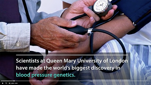
The video above summarizes research led by Queen Mary University of London and Imperial College London, which found over 500 new gene regions that influence people’s blood pressure, in the largest global genetic study of blood pressure to date. Click here to view the video. (Photo and caption copyright: Queen Mary University of London.)
Genetics Influence Blood Pressure More Than Previously Thought
In addition to identifying hundreds of new genetic regions influencing blood pressure, the researchers compared people with the highest genetic risk of high blood pressure to those in the low risk group. Based on this comparison, the researchers determined that all genetic variants were associated with:
- “having around a 13 mm Hg higher blood pressure;
- “having 3.34 times the odds for increased risk of hypertension; and,
- “1.52 times the odds for increased risk of poor cardiovascular outcomes.”
“We identify 535 novel blood pressure loci that not only offer new biological insights into blood pressure regulation, but also highlight shared genetic architecture between blood pressure and lifestyle exposures. Our findings identify new biological pathways for blood pressure regulation with potential for improved cardiovascular disease prevention in the future,” the researchers wrote in Nature Genetics.
Other Findings Link Known Genes and Drugs to Hypertension
The UK researchers also revealed the Apolipoprotein E (ApoE) gene’s relation to hypertension. This gene has been associated with both Alzheimer’s and coronary artery diseases, noted Lab Roots. The study also found that Canagliflozin, a drug used in type 2 diabetes treatment, could be repurposed to also address hypertension.
“Identifying genetic signals will increasingly help us to split patients into groups based on their risk of disease,” Paul Elliott, PhD, Professor, Imperial College London Faculty of Medicine, School of Public Health, and co-lead author, stated in the news release. “By identifying those patients who have the greatest underlying risk, we may be able to help them to change lifestyle factors which make them more likely to develop disease, as well as enabling doctors to provide them with targeted treatments earlier.”
Working to Advance Precision Medicine
The study shares new and important information about how genetics may influence blood pressure. By acquiring data from more than one million people, the UK researchers also may be setting a new expectation for research about diagnostic tests that could become part of the test menu at clinical laboratories throughout the world. The work could help physicians and patients understand risk of high blood pressure and how precision medicine and lifestyle changes can possibly work to prevent heart attacks and strokes among people worldwide.
—Donna Marie Pocius
Related Information:
Study of One Million People Leads to World’s Biggest Advance in Blood Pressure Genetics
Researchers Find 535 New Gene Regions That Influence Blood Pressure
Genetic Analysis of Over One Million Identifies 535 New Loci Associated with Blood Pressure Traits
The Facts About High Blood Pressure
High Blood Pressure Breakthrough: Over 500 Genes Uncovered
Study of a Million People Reveals Hypertension Genes


