Computer-aided diagnostic system combines optical dermatoscopy, spectrophotometry and high-frequency ultrasound imaging techniques to differentiate malignant lesions from benign moles
Detecting skin cancer via the use of skin biopsies is the bread and butter of many dermatopathology practices. But new technologies that can instantly detect and distinguish different types of skin malignancies may result in a reduced flow of skin biopsies to dermatopathologists in the not-too-distant future.
The new technique achieved a more accurate method of differentiating melanoma from benign lesions, according to the researchers.
“The novelty of our method is that it combines diagnostic information obtained from different non-invasive imaging technologies such as optical spectrophotometry and [high-frequency] ultrasound. Based on the results of our research, we can confirm that the developed automated system can complement the non-invasive diagnostic methods currently applied in the medical practice by efficiently differentiating melanoma from a melanocytic mole,” said Renaldas Raišutis, PhD, coauthor of the study, in a KTU news release.
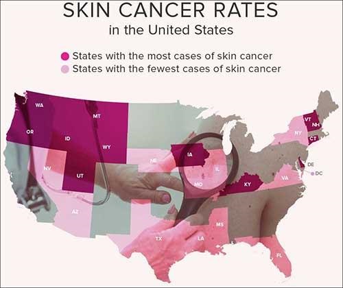
According to the Skin Cancer Foundation, one in five Americans will develop skin cancer by the age of 70 and more than two people in the United States die of skin cancer every hour. Early diagnosis is vital. If a malignant melanoma—the most lethal type of skin cancer—is found early, the five-year survival rate is 99%. (Graphic copyright: Healthline.)
“An efficient diagnosis of an early-stage malignant skin tumor could save critical time, more patients could be examined, and more of them could be saved,” Raišutis said in the news release. He added that the CADx-based diagnostic system is aimed at medical professionals but at a price that makes it affordable for smaller medical institutions. The Lithuanian team also is working to design a system that could be marketed for home use.
New Non-invasive Optical Technology May Reduce Demand for Skin Biopsies
A systematic review article published in Frontiers in Medicine Dermatology compared current diagnostic techniques for melanoma. It noted, “The current gold standard for melanoma diagnosis is the administration of dermoscopy, followed by a biopsy and subsequent histopathological analysis of the excised tissue. To minimize the risk of misdiagnosis of true melanomas, a significant number of dermoscopically ambiguous lesions are biopsied [increasing] the overall diagnostic costs and time to obtain the final diagnosis.”
But continuing technological innovations may be setting the stage for a reduction in the number of skin biopsies performed each year. In addition to the novel diagnostic method announced by the Lithuanian researchers, an Israeli scientist has created an innovative optical technology that can instantly and non-invasively detect and distinguish between three primary skin cancers:
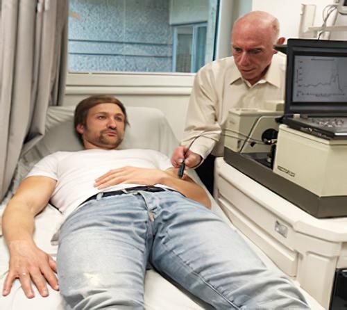
Tel Aviv University Physics Professor Abraham Katzir, PhD (above), demonstrates his new method of detecting cancerous skin lesion, which employs infrared sensors and optical fibers to determine the properties of various lesions on the skin and identify them based on their coloration within the infrared spectrum. (Photo copyright: Tel Aviv University.)
“We figured that with the help of devices that can identify these colors, healthy skin and each of the benign and malignant lesions would have different ‘colors,’ which would enable us to identify melanoma,” Katzir said in an Israel 21c news article.
“Melanoma is a life-threatening cancer, so it is very important to diagnose it early on, when it is still superficial,” Katzir told Israel 21c, adding that the new technology has the potential to cause “dramatic change” in the field of diagnosing and treating skin cancer, “and perhaps other types of cancer as well.”
As advancements in the non-invasive diagnosis of skin cancers continue, dermatopathologists—and in fact all anatomic and histopathology practices—should prepare for the financial impact this change may have on their clinical practices as demand for skin biopsies decreases.
Intriguing technology may find immediate value in assisting the detection and tracking of COVID-19 worldwide
Pathologists and clinical laboratory personnel old enough to have watched Star Trek on television will recall the tricorder, a multi-functional handheld device that could non-invasively detect any disease or medical condition that the science fiction series needed to be revealed. Fiction, yes, but so was the Star Trekcommunicator before the advent of smartphones.
Now, Florida-based Advanced Medical Solutions International (AMSI) anticipates bringing to market in early 2022 a similar tricorder-like handheld device that detects SARS-CoV-2 in humans and on contaminated objects and surfaces.
AMSI’s COVID Hunter™ device would be the world’s first noninvasive touchless viral detector for COVID-19, which has reportedly killed 4.55 million people worldwide. The inventors make the point that the device is simply to detect the presence of the coronavirus. It is not a diagnostic test.
For clinical laboratory scientists, this is yet another example of new technology being applied to a clinical problem that could ultimately lead to new diagnostic tools, not only for COVID-19, but ultimately for other viruses as well.
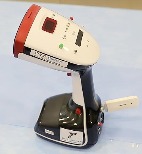
Pictured above is the actual COVID Hunter™ device that was extensively used in testing around the world. According to AMSI, this breakthrough technology can immediately detect COVID-19 in a person’s throat, lungs, sinuses, and breath, or on skin or clothes. High-touch areas such as door handles, mobile phones, and desktops also could be routinely checked for the virus and sanitized, breaking the transmission chain. (Photo copyright: Advanced Medical Solutions International.)
According to the COVID Hunter™ website, the device’s proprietary detection method utilizes a US-patent-pending detection technology that was initially invented by Engineer Nassar Said, a partner and inventor at AMSI. The method for detecting SARS-CoV-2 (the coronavirus that causes COVID-19) utilizes the above patent-pending detection technology and was invented and developed by Nassar Said and Adeeb Al-Zoubi, PhD, immunologist, and AMSI co-founder and Chief Scientific Officer.
According to the inventors, the detection technology employed by the COVID Hunter™ utilizes a combination of radio frequency (RF) and infrared (IR) electromagnetic waves to detect the RNA and spike protein found in the SARS-CoV-2 coronavirus with greater than 99% specificity and 99% sensitivity from as far as six feet away.
Al-Zoubi described the groundbreaking technology in a January 2021 news conference introducing the device. “This patent-pending technology uses a unique combination of light waves and sound waves combined to hone in on specific physical, chemical, and biological characteristics of SARS-CoV-2,” he said.
“We are basically surrounding the virus and characterizing the virus on all its characteristics all at once,” he continued. “Through focused research and tireless work, we at AMSI and Stem Cells Arabia [a Jordanian scientific research company] analyzed and specified these physical, chemical, and biological characteristics of SARS-CoV-2 and used these characteristics as one single value to target the detection by the COVID Hunter™.
“The sum of these specific SARS-CoV-2 characteristics is not found in any other virus or any other targets and constitutes a unique thumbprint of the virus,” he added.
“The handheld COVID Hunter™ will revolutionize the way SARS-CoV-2 (including mutated strains) is detected, slowing the spread of the deadly virus, saving lives, and returning life to ‘normal’ in the near future,” said AMSI co-founder and CEO Donald Redman (above center), with technology inventor/AMSI partner Nassar Said (left) and AMSI co-founder/Chief Scientific Officer and COVID Hunter™ co-inventor Adeeb Al-Zoubi, PhD (right), in a news release.
The COVID Hunter™ introductory press conference noted:
The COVID Hunter™ showed 100% accuracy and 100% specificity to detect only SARS-CoV-2 positive samples, distinguishing COVID-19 from viruses such as SARS-CoV-1, MERS, Influenza, and HIV,
The COVID Hunter™ detected all PCR positive COVID-19 test samples among more than 4,000 nasal swabs.
When more than 1,000 human subjects were tested with both PCR testing and the COVID Hunter™, the device confirmed as positive all confirmed COVID-19 cases.
4.8% of PCR false negatives in human subjects were accurately detected by the COVID Hunter™ as COVID-19 positive, indicating superior sensitivity to PCR testing.
76 out 94 confirmed COVID-19 positive individuals were shown to be infective, meaning they could transmit the disease.
The COVID Hunter™ was able to track the mode of transmission of COVID-19 as the virus moved from hand to mouth to other people and objects. Developers found that a healthy individual who shook hands with an infected person could transmit the virus to a third party without becoming infected themselves.
Researchers detected COVID-19 on the feet of domestic pets, indicating pets could transmit the virus to multiple persons within a household.
Al-Zoubi said nine months of research and development resulted in several COVID Hunter™ prototypes that demonstrated accuracy, specificity, and sensitivity in experiments using both nasal swab samples and confirmed COVID-19 patients residing in quarantine areas and hospitals in different countries.
“I am excited to see the COVID Hunter™ go from the prototype phase to a fully refined manufactured device that can be used to save lives around the world,” Al-Zoubi said in his concluding remarks.
Mass Production of COVID-Hunter
In an exclusive interview with Dark Daily, Redman and Al-Zoubi said they are seeking additional investor backing so they can shift from product refinement to high-volume manufacturing. If funding is secured this fall, their goal is to begin production in January 2022 of up to 30,000 units per month, which are projected to sell for $3,000 per device. Initially, the COVID Hunter™ would be marketed only as a COVID-19 detection tool under Federal Trade Commission (FTC) regulations.
Once manufacturing begins, AMSI will be able to submit the required number of COVID Hunter™ devices to the federal Food and Drug Administration (FDA) for review, the final step in its application for Emergency Use Authorization (EUA) of the COVID Hunter™ as a COVID-19 diagnostic device. The company expects its expedited EUA review to be completed by early spring.
AMSI notes that COVID Hunter™ can perform up to 300 scans per hour and does not use consumables other than batteries. This, according to Al-Zoubi, makes it a game-changing device for the travel industry, schools, businesses, restaurants, professional sports franchises, and concert venues seeking a return to “normal” operations.
The COVID Hunter™ also will be capable of being updated online to precisely detect new virus mutations, making it a critical weapon to defeat the pandemic as new COVID-19 mutations are found.
“This device is highly tested and it’s much more accurate than PCR [testing] because it detects the virus based on the physical presence of the virus, not based on chemical reactions or antibodies,” Al-Zoubi told Dark Daily. “We have gone beyond proof-of-concept testing.”
Clinical pathologists will want to follow development of the COVID Hunter™ and see if it eventually receives FDA approval. It may fulfill its promise as a game-changing new technology, not just for detection, but also for diagnosis.
The inventors and developers of the COVID Hunter™ will present their technology and its potential uses in detection and diagnosis at the upcoming Executive War College on Laboratory and Pathology Management, which takes place at the San Antonio Hyatt Riverwalk Hotel on Nov. 2-3, 2021.
Adeeb Al-Zoubi, PhD, and Nassar Said will conduct the session titled “New Technology Preview: Meet the COVID Hunter™, a Non-Invasive, Touchless, Immediate, and Portable Detection Device That Identifies the SARS-Cov-2 Virus.”
Medical laboratory professionals interested in attending this informative presentation can register by clicking here or by copying https://www.executivewarcollege.com your browser.
The ongoing study shows promise in the general development of self-powered wearable biosensors, the researchers say, in a development that has implications for clinical laboratory testing
Years back, it would be science fiction to describe a wearable garment that can not only measure an individual’s biomarkers in real-time, but also generates the power the device needs from the very specimen used for the measurement. Clinical laboratory managers and pathologists may find this new technology to be an interesting milestone on the path to wearable diagnostic devices.
With cases of diabetes on the rise across the globe, innovative ways to monitor the disease and simplify care is critical for effective diagnoses and treatment. Now, a team of researchers at Tokyo University of Science (TUS) in Japan have recently developed a diaper that detects blood glucose levels in individuals living with this debilitating illness.
Of equal interest, this glucose-testing diaper has a self-powered sensor that utilizes a biofuel cell to detect the presence of urine, measure its glucose concentration, and then wirelessly transmit that information to medical personnel and patients. The biofuel cell generates its own power directly from the urine.
Glucose in urine provides valuable data regarding blood sugar levels and can be used as an alternative to frequent blood draws to measure those levels. Monitoring the onset and progression of diabetes is crucial to making patient care easier, particularly in elderly and long-term care patients. Widespread use of these diapers in skilled nursing facilities and other healthcare settings could create an opportunity for clinical laboratories to do real-time monitoring of the blood sugar measurements and alert providers when a patient’s glucose levels indicate the need for attention.
“Besides monitoring glucose in the context of diabetes, diaper sensors can be used to remotely check for the presence of urine if you stock up on sugar as fuel in advance,” said Isao Shitanda, PhD, Associate Professor at the Department of Pure and Applied Chemistry, Faculty of Science and Technology, Tokyo University of Science, in a TUS press release. “In hospitals or nursing care sites, where potentially hundreds of diapers have to be checked periodically, the proposed device could take a great weight off the shoulders of caregivers,” he added.
Through electrochemistry, the scientists created their paper-based biofuel cell so that it could determine the amount of glucose in urine via reduction oxidation reactions, or redox for short. Using a process known as “graft polymerization,” they developed a special anode that allowed them to “anchor glucose-reactive enzymes and mediator molecules to a porous carbon layer, which served as the base conductive material,” the press release noted.
The biosensor was tested using artificial urine at different glucose levels. The energy generated from the urine then was used to power up a Bluetooth transmitter to remotely monitor the urine concentration via a smartphone. The TUS researchers determined their biofuel cell was able to detect sugar levels present in urine within one second. The diaper with its sensor could help provide reliable and easy monitoring for diabetic and pre-diabetic patients.
“We believe the concept developed in this study could become a very promising tool towards the general development of self-powered wearable biosensors,” Shitanda said in the press release.
According to the Isao Shitanda, PhD (above), lead author of the TUS study, 34.2 million people, or just over 10% of the US population, were diagnosed with diabetes in 2020. The federal Centers for Disease Control and Prevention estimates that an additional 7.3 million people have diabetes and are undiagnosed. A self-powered biosensor that detects diabetes and prediabetes in urine could help clinical laboratories and doctors catch the disease early and/or monitor its treatment. (Photo copyright: Tokyo University of Science.)
The World Health Organization (WHO) estimates that 422 million people globally were living with diabetes in 2014, and that 1.5 million deaths could be attributed directly to diabetes in 2019.
A panel of colored squares embedded on the front of the diaper changed color if specific chemical reactions fell outside normal parameters. If such a color change was observed, a smart phone application could relay that information to the baby’s doctor to determine if any further testing was needed.
Since we wrote that ebriefing in 2013, Pixie Scientific has expanded its product line to include Pixie Smart Pads, which when added to a diaper, enable’s caregivers to monitor wearers for urinary tract infections (UTI) and report findings by smartphone to their doctors.
These examples demonstrate ways in which scientists are working to combine diagnostics with existing products to help people better manage their health. Wearable electronics and biosensors are increasingly helping medical professionals and patients monitor bodily functions and chronic diseases.
As clever as these new wearable devices may be, there is still the need to monitor the diagnostic data they produce and interpret this data as appropriate to the patient’s state of health. Thus, it is likely that pathologists and clinical laboratory professionals will continue to play an important role in helping consumers and providers interpret diagnostic information collected by wearable, point-of-care testing technology.
If tattoos can accurately be used in the diagnostic process, might clinical laboratories soon offer these types of diagnostic tattoos at their patient service centers?
Could color-changing tattoos help diagnose illnesses? Researchers at the ATLAS Institute at the University of Colorado Boulder think so. They are working on prototypes of permanent tattoos that can detect chemical changes in the body and smart tattoo ink that would take the concept of wearable medical devices to a whole new level.
Called “dynamic” or “smart” tattoos, these color-changing tattoos have a biomedical purpose. They alert individuals to potential health issues due to changes in the biochemistry in their body. The technology has already been used in animal studies to detect sodium, glucose, electrolytes, and pH levels. Pathologists and clinical lab manager will recognize the value of a relatively non-invasive way to measure and track changes in these types of biomarkers.
“We developed a photochromic tattoo that serves as an intradermal ultraviolet (UV) radiometer that provides naked-eye feedback about UV exposure in real time. These small tattoos, or ‘solar freckles’, comprise dermally implanted colorimetric UV sensors in the form of nano encapsulated leuco dyes that become more blue in color with increasing UV irradiance,” the ATLAS scientists wrote.
Studies analyzing the efficacy of dynamic tattoos have provided strong evidence that they can be engineered to change color and sense and convey medical information. This field is called “dynamic tattoos” and in recent years various proof-of-concept studies have demonstrated that tattoos can be used to “pick up changes in sodium, glucose, electrolytes or pH levels in animal models,” Labroots reported.
“We demonstrate the tattoos’ functionality for both quantitative and naked-eye UV sensing in porcine skin ex vivo, as well as in human skin in vivo. Solar freckles offer an alternative and complementary approach to self-monitoring UV exposure for the sake of skin cancer prevention,” the researchers explained in their ACS Nano article.
“Activated solar freckles provide a visual reminder to protect the skin, and their color disappears rapidly upon removal of UV exposure or application of topical sunscreen. The sensors are implanted in a minimally invasive procedure that lasts only a few seconds yet remain functional for months to years,” they added.
“These semipermanent tattoos provide an early proof-of-concept for long-term intradermal sensing nanomaterials that provide users with biomedically relevant information in the form of an observable color change,” the ATLAS researchers concluded.
“When you think about what a tattoo is, it’s just a bunch of particles that sit in your skin,” Carson Bruns, PhD, Assistant Professor, Laboratory for Emergent Nanomaterials, ATLAS Institute, Mechanical Engineering, told Technology.org. “Our thought is: What if we use nanotechnology to give these particles some function?”
The invisible tattoos Bruns and the ATLAS team created turn blue in the presence of harmful levels of ultraviolet radiation to inform wearers that their skin needs protection and to apply or reapply sunscreen.
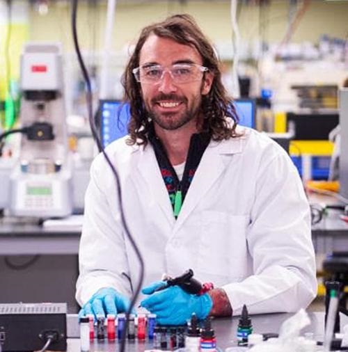
“I have always been interested in both art and science. My favorite type of art is tattooing and my favorite type of science is nanotechnology,” Carson Bruns, PhD (above), Assistant Professor, Mechanical Engineering, ATLAS Institute, told Inked. “When I had an opportunity to start a new research program, I thought it would be really fun and interesting to try and put the two together.” Might innovative medical laboratories one day operate “tattoo parlors” in their patient service centers to provide patients with tattoos that monitor key biometrics? (Photo copyright: Inked.)
The tattoo ink used for these tattoos contains a UV-activated dye inside of a plastic nano capsule that is less than a thousandth of a millimeter in size, or several sizes smaller than the width of a human hair. The capsules protect the dyes from wear and tear while allowing them to sense and respond to biochemical changes in the body. These tattoos are implanted into the skin using tattoo machines, much like getting a regular tattoo.
“I call them solar freckles because they’re like invisible freckles that are powered by sunshine,” Bruns told Inked, adding, “Millions of cases of preventable skin cancer are treated every year. I hope that the UV-sensitive tattoo will help us reduce the number of those cases by reminding people when their skin is exposed to unsafe levels of UV light.”
Dynamic Tattoos May Help People Lead Healthier Lives
One downside to these tattoos is that they only last a few months before they begin to degrade, requiring the wearer to get a “booster” tattoo.
The researchers hope that someday similar tattoo technologies will be applied to a wide variety of preventative and diagnostic applications. The goal is to enable people to detect health issues and allow them to lead healthier lives.
“We want to make tattoos that will allow you to, for example, sense things that you can’t currently sense,” Bruns told Inked. “Sometimes I joke that we want to make tattoos that give you superpowers.”
The ATLAS scientists imagine a future where tattoos can detect things like blood alcohol levels or high/low blood sugar levels or other changes in a person’s biochemistry.
“More generally, I hope that smart tattoos will help people stay healthy and more informed about their body, while also giving people new ways to express themselves creatively,” Bruns said.
Using Dynamic Tattoo to Detect Cancer
In 2018, a team of biologists created a tattoo comprised of engineered skin cells and an implantable sensor which could detect elevated blood calcium levels that are present in many types of cancers. These cancer-detecting tattoos were tested on living mice and would darken to notify researchers of potential problems.
Scientists at the Department of Biosystems Science and Engineering at the Swiss Federal Institute of Technology Zurich (ETHZ), Switzerland, developed a biomedical tattoo that uses bio sensitive ink and changes color based on variations in the body’s interstitial fluid. It recognizes four widespread cancers:
breast,
colon,
lung, and
prostate.
“Nowadays, people generally go to the doctor only when the tumor begins to cause problems. Unfortunately, by that point it is often too late,” Martin Fussenegger, PhD, Professor of Biotechnology and Bioengineering at the Department of Biosystems Science and Engineering (D-BSSE) of the ETH Zurich in Basel as well as at the University of Basel, told Medical News Today.
“For example, if breast cancer is detected early, the chance of recovery is 98%,” he continued. “However, if the tumor is diagnosed too late, only one in four women has a good chance of recovery.”
Though it appears that dynamic tattoos may be a functional and decorative way to track health, rigorous research and safety testing on human subjects will be required before clinical laboratories can set up diagnostic tattoo parlors in their offices.
Nevertheless, this concept demonstrates how different technologies under development may provide clinical laboratories with innovative and unusual diagnostic tools in the future.
Goal of university’s yearlong CHURP test was to validate the use of unmanned aircraft systems (UAS), commonly known as drones, in the delivery of medical supplies across SUNY’s campus
Just as hospital systems worldwide are exploring the feasibility of using drone technology to deliver clinical laboratory specimens and medical supplies between healthcare settings and medical laboratories, SUNY Upstate Medical University also has joined the growing list of healthcare providers that have added unmanned aerial vehicles (UAVs), or drones, to their specimen/supplies delivery services.
Traditional delivery of similar items normally takes about seven minutes. The drone delivered the same test kit in just two minutes, according to Government Technology(GT).
Then, “To prove that drone deliveries can be scaled up, the team conducted more medical deliveries in three locations throughout Syracuse two weeks ago, sending supplies from the hospital to a medical laboratory, from the hospital to a surgery center, and from a pharmacy to a second hospital,” GT reported.
Tony Basile, Chief Operations Officer at NUAIR, told GT the flight was a “proof of concept demonstration to show that medical deliveries can be made by drone when speed is essential, such as when tissue samples taken from a surgery patient must be delivered rapidly to a laboratory in a different building.”
Special FAA Waiver Allows Drone Flights Over Certain People
The FAA’s 107.39 waiver allows a drone operator to fly over people who are not participating in the operation and over those who are not covered under a structure or within a stationary vehicle. The January flight marked the first time the FAA’s 107.39 waiver was used for such a drone operation, a DroneUp press release notes.
The SUNY and the NUAIR alliance began formulating the concept of using drones to make medical deliveries more than a year and ago. At that time, there were concerns that a nearby highway project would disrupt normal clinical laboratory specimen delivery operations. The highway separates the hospital from a surgery center and finding a way to expedite deliveries despite slow traffic was essential, GT reported.
“They’re not going to want to wait 20 minutes for a tissue sample to get to the lab because the highway is coming down,” Basile told GT.
Challenges Encountered with Drone Delivery of Clinical Specimens and Supplies
In late spring, the team conducted additional deliveries to further prove the efficacy of using drones to transport medical supplies. They successfully transported supplies via UAV from the SUNY hospital to a clinical laboratory, from the hospital to a separate surgery center, and from a pharmacy to another hospital in the area.
“We conducted 52 successful deliveries throughout the week and were able to streamline the process, getting to about five deliveries an hour,” Basile wrote in a NUAIR article he penned, titled, “Making Drone Deliveries Scalable and Economically Viable.”
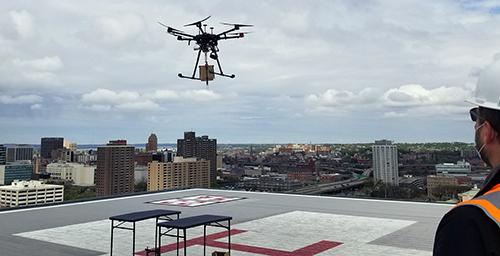
The drone used during the NUAIR test flights was equipped with a parachute in case of a mechanical failure and was only flown late at night. Basile believes the FAA will likely require medical delivery drones to have six to eight motors (shown above) as a security measure in case one or two motors fail. A common type of drone, known as a quadcopter, has only four motors and it is unable to fly if even one motor fails. (Photo copyright: NUAIR.)
Although the unmanned delivery flights were successful, SUNY experienced challenges with using drones to make medical deliveries. Those challenges included:
Economics: The NUAIR test flights required five people to conduct the flights, which is more costly than paying one driver to deliver the supplies.
FAA restrictions: The FAA currently does not allow biohazardous materials or controlled substances to be transported by drones due to the risk of public exposure if a crash occurs.
Device approval: The FAA is still in the process of evaluating which drone models will be permitted to carry medical supplies.
Weather: Drones cannot fly in inclement weather conditions.
“[Drones] are susceptible to strong winds and icing,” Basile told GT.
However, Basile believes that with more research and test flights the challenges will be resolved, and that drones will be used for medical deliveries in the future.
“I think they’re certainly going to be used,” he told GT. “Whether it’s soon depends on what you mean by soon.”
Drones Deliver Clinical Laboratory Specimens and Medical Supplies Worldwide
Other countries are increasingly using drones to deliver COVID-19 test kits and samples to and from remote areas.
In 2018, Dark Daily reported on automated logistics company Zipline’s use of fixed-wing drones called “Zips” to provide on-demand access to vital blood supplies in Rwanda and Tanzania. The Silicon Valley company transported more than 5,500 units of blood in 2017 to 12 regional hospitals from a base in the east of Rwanda, reported The Guardian. Zipline began operating in the African nation in 2016 and quickly cut blood delivery time from four hours to an average of about 30 minutes.
And in “Swiss Post Medical Drone Carrying Clinical Laboratory Specimens Crashes in Switzerland,” we reported that the medical drone revolution experienced a setback when drone-pioneer Swiss Post (Switzerland’s postal service) saw one of its American-made Matternet drones crash into Lake Zurich, Switzerland. According to a Swiss Post news release, the drone went down carrying a “non-vital” blood sample (one that had been previously analyzed). The flight was part of a recently launched pilot program transporting blood samples between Zurich’s central laboratory and the Hirslanden Klinik Im Park, a private clinic on the opposite side of Lake Zurich.
Although not all drone delivery flights end in success, these projects clearly demonstrate how safe and reliable drone delivery of medical supplies and clinical laboratory specimens could one day be beneficial to medical communities.
Such drone deliveries will likely help medical professionals expedite diagnoses and treatment options for patients, especially in remote areas where land transportation would be much less timely.
As the worldwide demand for histopathology services increases faster than the increase in the number of anatomic pathologist and histopathologists, a DP platform that suggests courses of treatments may be a boon to cancer diagnostics
Europe may become Ground Zero for the widespread adoption of whole-slide imaging (WSI), digital pathology (DP) workflow, and the use of image-analysis algorithms to make primary diagnoses of cancer. Several forward-looking histopathology laboratories in different European countries are moving swiftly to adopt these innovative technologies.
Clinical laboratories and anatomic pathology groups worldwide have watched digital pathology tools evolve into powerful diagnostic aids. And though not yet employed for primary diagnoses, thanks to artificial intelligence (AI) and machine learning many DP platforms are moving closer to daily clinical use and new collaborations with pathologists who utilize the technology to confirm cancer and other chronic diseases.
Now, Swiss company Unilabs, one of the largest laboratory, imaging, and pathology diagnostic developers in Europe, and Israel-based Ibex Medical Analytics, developer of AI-based digital pathology and cancer diagnostics, have teamed together to deploy “Ibex’s multi-tissue AI-powered Galen platform” across 16 European nations, according to a Unilabs press release.
Though not cleared by the federal Food and Drug Administration (FDA) for clinical use in the US, the FDA recently granted Breakthrough Device Designation to Ibex’s Galen platform. This designation is part of the FDA’s Breakthrough Device Program which was created to help expedite the development, assessment, and review of certain medical devices and products that promise to provide for more effective treatment or diagnosis of life-threatening or irreversibly debilitating diseases or conditions.
Benefits of AI-Digital Pathology to Pathologists, Clinical Labs, and Patients
According to Ibex’s website, the Galen DP platform uses AI algorithms to analyze images from breast and prostate tissue biopsies and provide insights that help pathologists and physicians determine the best treatment options for cancer patients.
This will, Ibex says, give pathologists “More time to dedicate to complex cases and research,” and will make reading biopsies “Less tedious, tiring, and stressful.”
Patients, according to Ibex, benefit from “Increased diagnostic accuracy” and “More objective results.”
And pathology laboratories benefit from “Increased efficiency, decreased turnaround time, and improved quality of service,” Ibex claims.
According to the press release, AI-generated insights can include “case prioritization worklists, cancer heatmaps, tumor grading and measurements, streamlined reporting tools and more.”
This more collaborative approach between pathologists and AI is a somewhat different use of digital pathology, which primarily has been used to confirm pathologists’ diagnoses, rather than helping to identify cancer and suggest courses of treatment to pathologists.
“This cutting-edge AI technology will help our teams quickly prioritize urgent cases, speed up diagnosis, and improve quality by adding an extra set of digital eyes,” said Christian Rebhan, MD, PhD (above), Chief Medical and Operations Officer at Unilabs, in the press release. “When it comes to cancer, the earlier you catch it, the better the prognosis—so getting us critical results faster will help save lives.” (Photo copyright: Unilabs.)
AI-based First and Second Reads
The utilization of the Galen platform will first be rolled out nationally in Sweden and then deployed in sixteen other countries. The AI-based DP platform is CE marked in the European Union for breast and prostate cancer detection in multiple workflows.
“The partnership with Ibex underlines Unilabs’ pioneering role in Digital Pathology and represents yet another step in our ambition to become the most digitally-enabled provider of diagnostic services in Europe,” Rebhan stated.
The Ibex website explains that the Galen platform is divided into two parts—First Read and Second Read:
The First Read “is an AI-based diagnostics application that aims to help pathologists significantly reduce turnaround time and improve diagnostic accuracy. The application uses a highly accurate AI algorithm to analyze slides prior to the pathologist and provides decision support tools that enable focusing on cancerous slides and areas of interest, streamline reporting, improve lab efficiency, and increase diagnostic confidence.”
The Second Read “is an AI-based diagnostics and quality control application that helps pathologists enhance diagnostic accuracy with no impact on routine workflow. The application analyzes slides in parallel with the pathologist and alerts in case of discrepancies with high clinical significance (e.g., a missed cancer), thereby providing a safety net that reduces error rates and enables a more efficient workflow.”
“Ibex is transforming cancer diagnosis with innovative AI solutions across the diagnostic pathway,” said Joseph Mossel, Chief Executive Officer and co-founder of Ibex, in the press release. “We are excited to partner with Unilabs to deploy our AI solutions and empower their pathologists with faster turnaround times and quality diagnosis. This cooperation follows a thorough evaluation of our technology at Unilabs and demonstrates the robustness and utility of our platform for everyday clinical practice.”
Use of AI in Pathology Increases as Number of Actual Pathologists Declines
Developers like Unilabs and Ibex believe that DP platforms driven by AI image analysis algorithms can help pathologists be more productive and can shorten the time it takes for physicians to make diagnoses and issue reports to patients.
This may be coming at a critical time. As nations around the globe face increasing shortages of pathologists and histopathologists, the use of AI in digital pathology could become more critical for disease diagnosis and treatment.
A 2019 Medscape survey stated that “One-third of active pathologists are burned out,” and that many pathologists are on the road to retirement.
And in the same year, Fierce Healthcare noted that in a 2013 study, “researchers found that more than 40% of pathologists were 55 or older. They predicted that retirements would reach their apex in 2021. Consequently, by the end of next decade, the United States will be short more than 5,700 pathologists.”
Dark Daily previously reported on the growing global shortage of pathologists going back to 2011.
Even China is struggling to keep up with demand for anatomic pathologists. In 2017, Dark Daily wrote, “China is currently facing a severe shortage of anatomic pathologists, which blocks patients’ access to quality care. The relatively small number of pathologists are often overworked, even as more patients want access to specialty care for illnesses. Some hospitals in China do not even have pathologists on staff. Thus, they rely on understaffed anatomic pathology departments at other facilities, or they use imaging only for diagnoses.”
Thus, it may be time for an AI-driven digital platform to arrive that can speed up and increase the accuracy of the cancer diagnostics process for pathologists, clinical laboratories, and patients alike.
There are multiple companies rapidly developing AI, machine learning, and image analysis products for diagnosing diseases. Pathologists should expect progress in this field to be ongoing and new capabilities regularly introduced into the market.