Thorough hand-washing protocols aren’t just for healthcare professionals anymore. Patients also need to be educated to prevent hospital-acquired infections
Microbiologists and clinical laboratory managers will be particularly interested to learn that patients are bringing deadly organisms into hospitals on their hands. That’s the conclusion of a University of Michigan (UM) study which found that as patients enter and move throughout hospitals, they deposit and spread multi-drug resistant organisms, or MDROs on clinical surfaces. When those surfaces are not properly decontaminated, the bacterial contamination spreads on contact.
This finding has implications for the nosocomial infection teams in hospitals that include microbiologists and clinical laboratories. After all, every day there is a large flow of walk-in patients and visitors who come in contact with dozens of surfaces. The potential for contamination with multi-drug resistant organisms is high.
Antibiotic-resistant bacteria have been the root cause of a marked increase in hospital-acquired infections (HAIs), which Dark Daily has covered extensively. That’s why healthcare professionals practice proper hand-washing protocols to help reduce the transmission of pathogens and curtail possible infections.
The UM study, however, suggests that patients also should be
educated on proper hand hygiene to diminish the potential spread of bacteria,
especially before making trips to the emergency room.
Between February and July of 2017, UM researchers at two
hospitals in Southeast Michigan tested 399 general medicine hospital patients
for the presence of MDROs, also known as superbugs. They swabbed the palms,
fingers, and around the nails of the patients’ dominant hands and the interior
of both nostrils.
The researchers found that 14% of the patients tested
positive for MDROs. In addition, nearly one third of high-touch objects and
surfaces in the hospital rooms tested positive for superbugs as well.
The hospital room surfaces that were swabbed for the
presence of MDROs were:
Due to the overuse of antibiotics, these types of bacteria
are often resistant to the drugs that were once used to kill them.
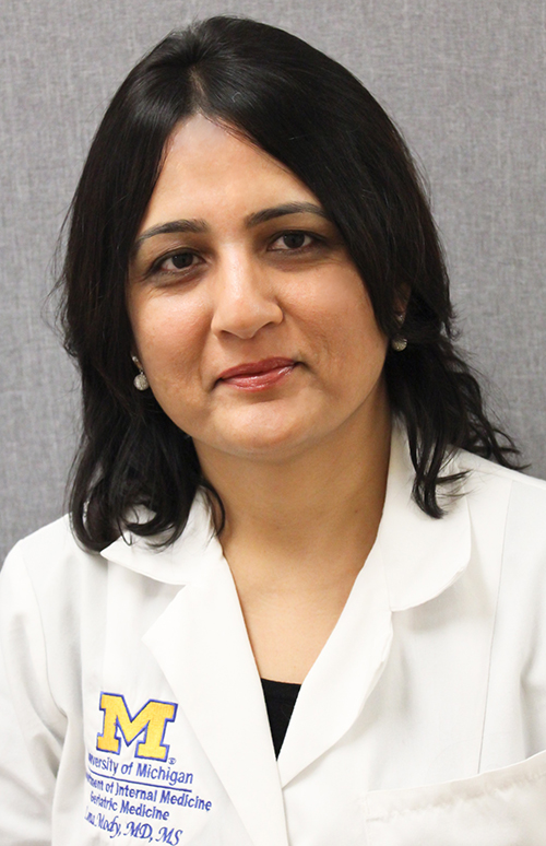
“Hand hygiene narrative has largely focused on physicians, nurses, and other frontline staff, and all the policies and performance measurements have centered on them, and rightfully so,” said Lona Mody, MD (above) in a press release. Mody is Professor of Internal Medicine at UM and one of the lead researchers for the study. “But our findings make an argument for addressing transmission of MDROs in a way that involves patients, too.”
Anatomy of a Hospital-Acquired Infection
The scientists tested patients and surfaces at different
stages of their hospital stays. The samples were taken on the day of admission,
days three and seven of the stays, and weekly thereafter until the patients
were discharged.
The team found that 6% of the patients who did not have
MDROs present at the beginning of their hospital stays tested positive for
superbugs at later stages of their stays. Additionally, 20% of the tested
objects and surfaces in the patients’ rooms had superbugs on them at later test
stages that were not present earlier in the hospital stays.
“This study highlights the importance of hand washing and environmental cleaning, especially within a healthcare setting where patients’ immune systems are compromised,” noted Katherine Reyes, MD, Department of Infectious Diseases, Henry Ford Hospital, in the press release. “This step is crucial not only for healthcare providers, but also for patients and their families. Germs are on our hands; you do not need to see to believe it. And they travel. When these germs are not washed off, they pass easily from person to person and objects to person and make people sick.”
Patients included in the study had to be new admissions, on
general medicine floors, and at least 18 years of age. Criteria that excluded
individuals from participation in the research included:
Being in observation status, typically after a
medical procedure;
Transfers from other hospitals;
Transfers from intensive care units;
Having cystic fibrosis (these patients have a
higher likelihood of MDRO colonization);
Receiving end-of-life care; and
Non-English speaking.
Patients who were transferred to a room on a
nonparticipating floor within the hospitals were immediately discharged from
the study.
Patients Travel Throughout Hospitals Spreading Germs
The presence of superbugs on patients or surfaces does not
automatically translate to a patient getting sick with antibiotic-resistant
bacteria. Only six of the patients in this study developed MRSA. However, all
six of those individuals tested positive for the superbug either on their hands
or on surfaces within their room.
The researchers noted that hospital patients typically do
not stay in their rooms. They are encouraged to walk throughout the hospital to
speed up the recovery process, and often are transported to other areas of
hospitals for medical tests and procedures. Patients also may be picking up
superbugs from other patients and staff members, other hospital areas, and
commonly-touched surfaces.
The UM researchers concluded in their study that “while the
burden of preventing infections has largely been borne by [healthcare
personnel], our study shows that patient hands are an important reservoir and
play a crucial role in the transmission of pathogens in acute care hospitals.
Thus, patient hand hygiene protocols should be implemented and tested for their
ability to reduce environmental contamination, pathogen transmission, and
healthcare-associated infections, as well as to increase meaningful patient
engagement in infection prevention.”
“Infection prevention is everybody’s business,” stated Mody
in the press release. “We are all in this together. No matter where you are, in
a healthcare environment or not, this study is a good reminder to clean your
hands often, using good techniques—especially before and after preparing food,
before eating food, after using a toilet, and before and after caring for
someone who is sick—to protect yourself and others.”
These
research findings should prove to be valuable for infection control teams and
microbiology laboratories in the nation’s hospitals and health systems, as well
as independent clinical laboratories, urgent care centers, and retail
healthcare clinics.
Learning
more about the transmission of infectious agents from patient to patient and
from surfaces to patients could aid in the development of new techniques and
strategies to prevent superbugs from manifesting in medical environments.
Miniaturization of clinical laboratory testing continues to intrigue pathology researchers, medical scientists, and diagnostics developers who see the technology as a way to bring pathology diagnostics to resource deficient areas
Can useful, fast, and cheap medical laboratory tests be performed using the million-pixel cameras found in today’s smartphones, in combination with microchips and other technologies? A team of researchers at Princeton University believe they are on the path to achieving those goals.
Dark Daily has covered the development of “lab-on-a-chip” miniature diagnostic technologies for many years. Through these diminutive devices, clinical laboratory testing has been brought to remote regions of the world where even basic resources like electricity and adequate clean water are in short supply.
The Princeton researchers are developing their own tiny biosensor microchip. The device reads fluorescent light and could, they say, be used to diagnose disease from inside the human body.
Revolutionary Use of Standard Microchip Technology
The device developed by the Princeton University researchers
uses silicon chip technology to perform various types of clinical laboratory
assays.
“The key idea is to allow complex optical systems in modern-day chips,” said Kaushik Sengupta, PhD, Assistant Professor of Electrical Engineering at Princeton and one of the project leaders, in a press release. “All smartphones carry a million-pixel camera. How do we turn this into a device that allows laboratory-quality diagnostics?”
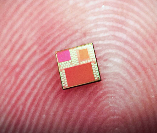
The miniature device (above) uses standard microchip technology consisting of tiny metal layers. It’s those layers that serve as the biosensor. The chip measures a mere four millimeters (approximately 5/32 of an inch) per side, and according to the University of Princeton scientists, it can be mass produced in a cost-effective manner using standard manufacturing techniques and does not require detailed assembly. (Photo copyright: Lingyu Hong/University of Princeton.)
The researchers discovered that existing microchip technology can be adapted to “take advantage of light’s unusual behavior when interacting with structures smaller than wavelength of light,” the press release noted.
“We show these complex optical biosensor systems can also be
realized in the same technology with absolutely no change in manufacturing the
microchip,” Sengupta said.
Employing existing manufacturing would make mass producing
the chips highly cost effective compared to other lab-on-a-chip technologies.
And, if the diagnostics are accurate as well, clinical laboratories could have
a remarkable new tool to aid physicians in the diagnosis of disease.
How It Works
The Princeton scientists say light harnessed by the fluorescence-based biosensor can detect and
differentiate biological substances ranging from bacterial Deoxyribonucleic acid (DNA)
to hormones present in humans.
They also claim their sensor can detect tiny molecules, such
as DNA and proteins, in liquid samples as small as one microliter. By
comparison, a single drop of water holds about 50 microliters. The researchers
say the sensitivity of their microchip in analyzing this tiny sample is
comparable to results achieve by diagnostic laboratories.
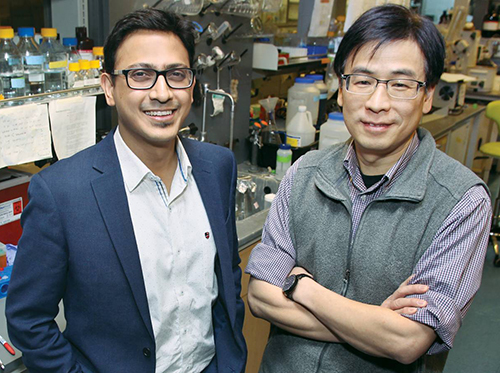
“We show for the first time that this level of optical field manipulation is possible in a silicon chip. By eliminating all classical optics, the system is now small enough that you could start thinking about putting it in a pill,” said Kaushik Sengupta, PhD, Assistant Professor of Electrical Engineering at Princeton. He’s shown above with Haw Yang, PhD (on right), Professor of Chemistry and Principle Investigator at the Haw Yang Laboratory at Princeton University. “You could start thinking about diagnostics inside the body in a way you could not think about before,” Sengupta concluded. (Photo copyright: Frank Wojciechowski/Princeton University.)
Like a traditional lab setup, the chip uses chemical
antibodies to target certain molecules. These antibodies are then altered to
propagate a specific light wavelength when they are exposed to a distinct
molecule. Exposure to ultraviolet light causes the antibodies to glow a faint
red color when they come into contact with the targeted substance.
Cheaper Diagnostics for the Developing World
The researchers hope that their miniature chip will someday
be used as a mainstream diagnostic technology, and that it may lead to the
development of other, similar diagnostic products.
“Once
we make the diagnostics cheaper, we can enable diagnostics in the developing
world,” stated Sengupta. “And it’s not just diagnostics. What we have come up
with here is just a low-cost, tiny fluorescent sensor and you can use
fluorescent sensing in many different things: for food and water-quality monitoring,
environmental monitoring, and industrial applications.”
More research is required to ensure the effectiveness of the
new technology. And it will need to receive clearance from the federal Food and Drug Administration (FDA) before going
into widespread production. Nevertheless, this newest miniature lab-on-a-chip
technology could prove beneficial to clinical laboratories in the future, as a
cost-effective tool to diagnose disease and better serve medical professionals
and patients in resource-strapped regions of the world.
Could clinical laboratories use texting to improving patient compliance with the medical laboratory test orders given to them by their doctors?
California’s largest physician-owned medical practice has
employed text messaging to reduce patient no-shows. Just as other innovations such
as same-day walk-in clinical laboratory
testing and patient at-home self-testing made it easier for patients to comply
with physicians’ lab test orders, text messaging appears to help get more
patients through the doors and into doctors’ exam rooms.
At least that’s the experience at Riverside Medical Clinic
(RMC) in Riverside, Calif. The multi-specialty practice has more than 170
providers who see more than 400,000 patients annually. After struggling to
lower its 15% baseline no-show rate using a phone-only reminder system, RMC turned
to a two-way texting appointment reminder system from Santa Barbara, Calif.-based
WELL Health (WELL).
According to a case
study, prior to the texting
system implementation, no-shows were costing RMC more than $3 million per year.
“The problem we were trying to resolve was getting a hold of our
patients in an expedient manner without having to do redundant work,” Diego
Galvez-Ramirez, Associate Vice President, Patient Business Services at
Riverside Medical Clinic, told Healthcare IT News. “We wanted to
give time back to our staff. A big frustration was not having enough time for
staff to accomplish their duties.”
After RMC implemented WELL’s HIPAA-compliant text-based reminder
system, front office efficiency and productivity improved, and the practice
experienced a 33% decrease in appointment no-shows.
Additionally:
No-shows decreased from 15% to 10% within the
first month of going live across the enterprise.
Confirmed appointments rose from 29.45% to
94.45%, translating to a savings of more than $40,000 in two months.
91% of patients who confirmed via WELL presented
for their visit.
Phone volume at RMC’s two call centers decreased
by 4% to 6%.
Galvez-Ramirez suggests that healthcare providers—including
clinical laboratories and anatomic pathology groups—keep pace with the
realities of today’s connected world. “Most of the time, the cell phone is not
used to make phone calls,” he told Healthcare IT News. “You have to adapt
to the new ways that your patients want and are used to communicating.
“In our environment,” he continued, “you also have to be
quick to respond to your patients. No patient wants to spend unnecessary time
on a phone call. Being able to send them their appointment to their phone is
not a new concept, it’s an expectation.”
Based on an Axway survey of 1,200 smartphone users aged 18-60, the graphic above supports the view that text messaging is now the preferred method of communications for most people. Could clinical laboratories employ text messaging to lower patient no-shows and increase the proportion of patients who actually show up at a patient service center to provide a specimen in response to the medical laboratory test orders given to them by their physicians? (Graphic copyright: MakingCharts.com/Axway.)
The WELL messaging app draws a patient’s information from the
physician’s electronic
health record (EHR) system to configure the appointment reminder. This
includes appointment type, date/time, and location. Based on the patient’s
preferred method, the system sends reminder messages via phone, text, or e-mail.
As Healthcare IT News noted, WELL’s competitors in the
patient communication space include:
Texting Reduces No-Shows at Other Healthcare Networks
Other healthcare organizations also have replicated RMC’s
success in reducing its no-show rates by moving away from telephone-based
reminders.
An Athena Health
study examined 54.3 million patient visits in 2015 and found no-show rates
dropped to 4.4% when patients received a reminder text from their provider. By
comparison:
Athena patients who received a phone call
instead of a text failed to show up 9.4% of the time;
E-mail reminders resulted in a 5.9% no-show rate;
and,
10.5% of patients who received no form of
reminder message missed their appointments.
Is Texting Secure and HIPAA Compliant?
A 2018 poll conducted by the Medical
Group Management Association (MGMA) found that 68% of healthcare organizations
used text messaging to communicate with patients about appointments. But is it
secure?
An MGMA
article notes that according to HIPAA Journal,
“Recent changes to HIPAA
have introduced new rules relating to how Protected
Health Information (PHI) should be communicated and many healthcare
organizations and other covered entities are now at risk of financial sanctions
and legal action should an avoidable breach of PHI occur.” The MGMA goes on to
state that, “As text messaging is not typically a fully-secure channel for the
communication of PHI, practices must be vigilant when sending information via
text messages.”
With proper training and precautions, clinical laboratories and
pathology groups might want to add text messaging to their patient outreach
programs. Data indicate that doing so could improve patient compliance with the
medical lab test orders given to them by their physicians. Industry experts
estimate that for every 100 medical lab test requests written by providers,
only about 60% of patients show up to provide the specimens needed for a lab to
perform those tests. Improving on those numbers would help clinical
laboratories and patients alike.
Scientists
at St. Jude’s have discovered that performing different genetic tests on pediatric
cancer patients, and then combining those test results, may help guide and
improve patient care.
The research was part of a St. Jude’s project called Genomes
for Kids (G4K), a study to determine how genetic information may be used to
diagnose and treat pediatric cancers.
Through this project, the researchers hope to learn why tumors form in
children and predict how tumors will respond to certain treatments.
‘It’s
a Whole Lot of Sequencing.’
Few tragedies are worse than cancer in children. This is where precision medicine treatments can be critical, and multiomics may play an important role in the development of new therapies.
Multiomics refers to a biological analysis approach in which
multiple “omes” are analyzed together in a collaborative way to locate relevant
biomarkers and functional relationships. These “omes” include:
To perform their research, the St. Jude scientists examined
253 pediatric cancer patients by conducting whole genome
sequencing (WGS), whole
exome sequencing (WES), and RNA
sequencing of their tumors. They also looked at the WGS and WES of
non-cancerous tissues extracted from the same cancer patients.
“It is a whole lot of sequencing. I admit that,” Scott Newman, PhD,
Group Lead, Bioinformatics Analysis at St. Jude’s, told The
Scientist.
“With results available in a clinically relevant time frame, and pricing becoming increasingly comparable to the radiology and pathology tests, WGS is becoming more accessible to pediatric oncology patients,” said Scott Newman, PhD (above), Group Lead, Bioinformatics Analysis, at St. Jude’s, in an American Society of Human Genetics (ASHG) news release. (Photo copyright: ASHG.)
As a result of their three-platform testing, the researchers
discovered there was at least one finding for each patient that could be useful
in providing a diagnosis, revealing risks for individual patients, or
pinpointing which drugs may be most beneficial for a particular patient in
nearly 200 (79%) of the cases. Such findings are at the heart of precision
medicine.
The researchers also compared their sequencing results to
cancer panels that use next-generation
sequencing (NGS) to target specific genes or mutations relevant to a
certain cancer phenotype.
During this portion of the research, they discovered that the cancer panels
missed 11% to 16% of actionable genes relating to diagnosis, prognosis, and
treatment.
“This is either good news or bad news, depending on how you
look at it,” Newman said. “Personally, I am amazed at how well these panels do
and how well they have been designed. But, if you want to know every mutation
that you would probably want to report, you have to do comprehensive
sequencing.”
First Multi-Platform Genomic Sequencing Study
“To
our knowledge, this is the first clinical study where this comprehensive three-platform
genomic sequencing approach was offered prospectively to all pediatric oncology
patients,” said Kim Nichols, MD,
Director, Division of Cancer Predisposition at St. Jude’s, in a St.
Jude’s blog post.
The testing costs $8,600 per patient, but is considered worth
it to improve patient diagnosis, prognosis, and treatment for pediatric cancer
patients.
“Compared with the cost of many
other procedures that children with cancer undergo, the cost is likely
comparable, or even less—for example, compared with complex surgical procedures
or multiple radiology tests,” Nichols said.
In addition, the test results are available in less than 30
days, which makes them more valuable, as time can be a critical asset to cancer
management.
The scientists hope this type of three-platform genetic
testing can help guide care for pediatric cancer patients.
“Because
so few of the molecular lesions in pediatric cancer are targetable by specific
drugs, currently it is the diagnostic and prognostic insights provided by the
three-platform approach that appear most clinically impactful,” said Nichols.
“From a diagnostic perspective, tumors may look the same under a microscope,
but the identification of specific genetic changes can direct you to the correct
diagnosis, and therefore, the most appropriate therapy. From a prognostic
perspective, you will have different risk stratifications depending on results.”
The results of the research were presented at the 2018
annual meeting of the American Society of Human Genetics in San Diego last
October. The St. Jude’s researchers hope that this type of research can drive
wider adoption of WGS in the assessment of pediatric tumors to improve patient
outcomes. Pathologists and medical laboratory scientists will want to watch for
additional research findings as the team at St. Jude’s uses this approach on
more pediatric cancer patients.
Methods that target the causes of acidity could become part of precision medicine cancer treatments and therapies
Researchers at Massachusetts
Institute of Technology (MIT) have found that acidic environments enable
tumor cells to strengthen through protein production. And that when acidic surfaces
extend beyond a tumor’s interior, and come into contact with healthy tissue,
cancer can spread.
The results of their study will interest anatomic
pathologists who review tissue biopsies to diagnose cancer and help identify
the most effective therapies for cancer patients. Currently, there are no new clinical
laboratory tests under development based on MIT’s research.
The researchers published their findings in the journal Cancer Research. Their paper also
shared how tumor acidity can be identified and reversed.
“Our findings reinforce the view that tumor acidification is an important driver of aggressive tumor phenotypes, and it indicates that methods that target this acidity could be of value therapeutically,” noted Frank Gertler, PhD (above), in a news release. Gertler is an MIT Professor of Biology, a member of MIT’s Koch Institute for Integrative Cancer Research, and a Senior Author of the study. (Photo copyright: MIT News.)
Acidity is a Tumor Cell’s
Friend
Acidity results from lack of oxygen in tumors and enables
tumor cell growth. “Acidification of the microenvironment plays established roles
in tumor progression and provides a hostile milieu that advantages tumor cell
survival and growth compared to non-cancerous cells,” the researchers wrote in Cancer Research.
In their study, the MIT scientists sought to learn:
What areas of a tumor are actually acidic?
How does acidosis propel cells to
invade surrounding healthy tissues?
They used a nanotechnology platform
called pHLIP (pH Low
Insertion Peptide) to sense pH at the surface of cancer cells and then insert a
molecular probe into the cell membranes. “This brings nanomaterial to close
proximity of cellular membrane,” noted a research study
conducted at the University of Rhode Island by scientists who developed the
pHLIP technology.
Medical News Today reported that the MIT scientists
used pHLIP to map the acidity in human breast cancer tumors implanted in mice.
When it detected a cell in an acidic environment, pHLIP sent a small protein
molecule into the cell’s membrane. The scientists found that acidosis was not
confined to the oxygen-rich tumor core. It extended to the stroma, an important boundary
between healthy tissue and malignant tumor cells.
“We characterized the spatial characteristics of acidic
tumor microenvironments using pHLIP technology, and demonstrated that
tumor-stroma interfaces are acidic, and that cells within the acidic front are
invasive and proliferative,” the scientists wrote in Cancer Research.
What Stimulates
Acidity and How to Reverse It?
The MIT researchers sought the reasons, beyond hypoxia, for
high acidity in tumor tissue.
“There was a great deal of tumor tissue that did not have
any hallmarks of hypoxia that was quite clearly exposed to acidosis. We started
looking at that, and we realized hypoxia probably wouldn’t explain the majority
of regions of the tumor that were acidic,” Gertler pointed out in the MIT news
release.
So what did explain it? The researchers pointed to aerobic
glycolysis, a “condition in which glucose is converted to lactate in the presence
of oxygen,” according to an article published by StatPearls. “Cancer
stem cells (CSC) within a tumor are notorious for aerobic glycolysis. Thus,
extensive aerobic glycolysis has been indicative of aggressive cancer,” the
paper’s authors noted.
During their study, the MIT scientists found:
Cells at the tumor surface shifted to aerobic
glycolysis, “a type of metabolism that generates lactic acid, making way
for high acidity,” and
“Tumor acidosis gives rise to the expression of molecules
involved in cell invasion and migration. This reprogramming, which is an
intracellular response to a drop in extracellular pH, gives the cancer cells
the ability to survive under low-pH conditions and proliferate,” said Nazanin Rohani, PhD, former
postdoctoral researcher in the MIT Koch Institute for Integrative Cancer
Research, and Lead Author of the study, in the news release.
Could a Reduction in Acidity Reverse Tumor Growth?
In another experiment, the researchers fed sodium
bicarbonate (baking soda) to mice with breast or lung tumors. The tumors became
less acidic and metastatic.
“It adds to the sense that this pH dynamic is not permanent.
It’s reversible. I think that’s an important addition to an ongoing discussion
about the role of pH in tumor behavior,” said Ian Robey, PhD, in an
MITblog
post. Robey is a Research Assistant Professor, Department of Medicine
at the University of Arizona, and Full Investigator at the Arizona Cancer Center. He was not
involved in the MIT research.
Spreading the Word on
How Cancer Spreads
The MIT study is important—not only to anatomic pathologists—but
also to oncologists and cancer patients worldwide. Cancer is not simple to
diagnose and treat. The MIT study may provide important insights into targeting
cancer care and precision
medicine treatments.
This research could lead to a useful liquid biopsy test that would be a powerful new tool for clinical laboratories and anatomic pathologists
Cancer researchers have long sought the Holy Grail of
diagnostics—a single biomarker that can quickly detect cancer from blood or
biopsied tissue. Now, researchers in Australia may have found that treasure. And
the preliminary diagnostic test they have developed reportedly can return
results in just 10 minutes with 90% accuracy.
In a news release, University of Queensland researchers discussed identifying a “simple signature” that was common to all forms of cancer, but which would stand out among healthy cells. This development will be of interest to both surgical pathologists and clinical laboratory managers. Many researchers looking for cancer markers in blood are using the term “liquid biopsies” to describe assays they hope to develop which would be less invasive than a tissue biopsy.
“This unique nano-scaled DNA signature appeared in every type of breast cancer we examined, and in other forms of cancer including prostate, colorectal, and lymphoma,” said Abu Sina, PhD, Postdoctoral Research Fellow at the Australian Institute for Bioengineering and Nanotechnology (AIBN), University of Queensland (UQ), in the news release.
“We designed a simple test using gold nanoparticles that
instantly change color to determine if the three-dimensional nanostructures of cancer
DNA are present,’ said Matt
Trau, PhD, Professor of Chemistry at the University of Queensland, and
Deputy Director and Co-Founder of UQ’s AIBN, in the news release.
The team’s test is preliminary, and more research is needed before
it will be ready for Australia’s histopathology laboratories (anatomic
pathology labs in the US). Still, UQ’s research is the latest example of how
increased knowledge of DNA is making it possible for researchers to identify
new biomarkers for cancer and other diseases.
“We certainly don’t know yet whether it’s the holy grail for
all cancer diagnostics, but it looks really interesting as an incredibly simple
universal marker of cancer, and as an accessible and inexpensive technology
that doesn’t require complicated lab-based equipment like DNA sequencing,” Trau
added.
The UQ researchers published their study in the journal Nature Communications. In it, they noted that “Epigenetic reprogramming in cancer genomes creates a distinct methylation landscape encompassing clustered methylation at regulatory regions separated by large intergenic tracks of hypomethylated regions. This methylation landscape that we referred to as ‘Methylscape’ is displayed by most cancer types, thus may serve as a universal cancer biomarker.”
While methyl patterning is not new, the UQ researchers say they were the first to note the effects of methyl pattern in a particular solution—water. With the aid of transmission electron microscopy, the scientists saw DNA fragments in three-dimensional structures in the water. But they did not observe the signature in normal tissues in water.
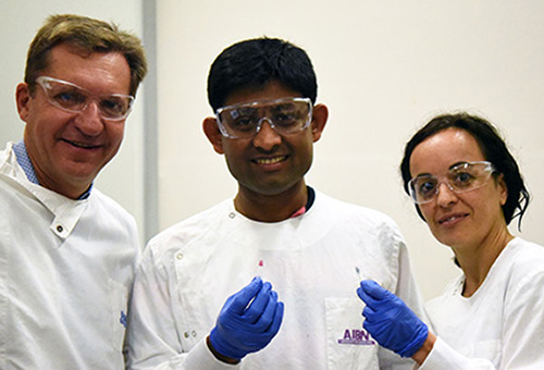
“To date, most research has focused on the biological consequences of DNA Methylscape changes, whereas its impact on DNA physicochemical properties remains unexplored,” UQ scientists Matt Trau, PhD (left), Abu Sina, PhD (center), and Laura Carrascosa (right), wrote in their study. “We exploit these Methylscape differences to develop simple, highly sensitive, and selective electrochemical or colorimetric one-step assays for the detection of cancer.” (Photo copyright: University of Queensland.)
Their test averaged 90% accuracy during the testing of 200
human cancer samples. Furthermore, the researchers found the DNA structure to
be the same in breast, prostate, and bowel cancers, as well as lymphomas, noted
The Conversation.
“We find that DNA polymeric
behavior is strongly affected by differential patterning of methylcytosine
leading to fundamental differences in DNA solvation and DNA-gold affinity
between cancerous and normal genomes,” the researchers wrote in NatureCommunications.“We exploit
these methylscape differences to develop simple, highly sensitive, and
selective electrochemical or one-step assays for detection of cancer.”
Next Steps for the
“Gold Test”
“This approach represents an exciting step forward in
detecting tumor DNA in blood samples and opens up the possibility of a generalized
blood-based test to detect cancer, Ged Brady, PhD, Cancer Research UK
Manchester Institute, told The
Oxford Scientist. “Further clinical studies are required to evaluate
the full clinic potential of the method.”
Researchers said the next step is a larger clinical study to
explore just how fast cancer can be detected. They expressed interest in
finding different cancers in body fluids and at various stages. Another opportunity
they envision is to use the cancer assay with a mobile device.
DiCarlo told USA Today
that such a mobile test could be helpful to clinicians needing fast answers for
people in rural areas. However, he’s also concerned about false positives. “You
don’t expect all tumors to have the same methylation pattern because there’s so
many different ways that cancer can develop,” he told USA Today. “There
are some pieces that don’t exactly align logically.”
The UQ researchers have produced an intriguing study that differs
from other liquid biopsy papers covered by Dark Daily. While their test may need to be used in combination with other
diagnostic tests—MRI, mammography, etc.—it has the potential to one day be used
by clinical laboratories to quickly reveal diverse types of cancers.