Labcorp, the commercial laboratory giant headquartered in Burlington, N.C., has billions of diagnostic test results archived. It takes samplings of those results and runs them through a machine learning algorithm that compares the data against a condition of interest, such as chronic kidney disease (CKD). Machine learning is a subdiscipline of AI.
Based on patterns it identifies, the machine learning algorithm can predict future test results for CKD based on patients’ testing histories, explained Stan Letovsky, PhD, Vice President for AI, Data Sciences, and Bioinformatics at Labcorp. Labcorp has found the accuracy of those predictions to be better than 90%, he added.
Labcorp also has created an AI-powered dashboard that—once layered over an electronic health record (EHR) system—allows physicians to configure views of an individual patient’s existing health data and add a predictive view based on the machine learning results.
For anatomic pathologists, this type of setup can quickly bring a trove of data into their hands, allowing them to be more efficient with patient diagnoses. The long-term implications of using this technology are significant for pathology groups’ bottom line.
Stan Letovsky, PhD (above), Vice President for AI, Data Sciences, and Bioinformatics at Labcorp, discussed AI developments in digital pathology during his keynote address at the 2022 Executive War College in New Orleans. “The best thing as a community that we can do for patients and their physicians with AI is to identify care gaps early on,” he said, adding, “If pathologists want to grow and improve their revenue, they have to be more productive.” (Photo copyright: Dark Intelligence Group).
Mayo Clinic Plans to Digitize 25 Million Glass Slides
In other AI developments, Mayo Clinic in Rochester, Minn., has started a project to digitally scan 25 million tissue samples on glass slides—some more than 100 years old. As part of the initiative, Mayo wants to digitize five million of those slides within three years and put them on the cloud, said pathologist and physician scientist Jason Hipp, MD, PhD, Chair of Computational Pathology and AI at Mayo Clinic.
“We want to be a hub within Mayo Clinic for digital pathology,” Hipp told Executive War College attendees during his keynote address.
Hipp views his team as the bridge between pathologists and the data science engineers who develop AI algorithms. Both sides must collaborate to move AI forward, he commented, yet most clinical laboratories and pathology groups have not yet developed those relationships.
“We want to embed both sides,” Hipp added. “We need the data scientists working with the pathologists side by side. That practical part is missing today.”
The future medical laboratory at Mayo Clinic will feature an intersection of pathology, computer technology, and patient data. Cloud storage is a big part of that vision.
“AI requires storage and lots of data to be practical,” Hipp said.
High court decision in 2012 altered patent law and effectively blocked protections for certain clinical laboratory diagnostic tests and procedures
Clinical laboratory leaders and pathologists will be interested to learn that a US Supreme Court (SCOTUS) decision from 2012 may be partly to blame for the shortage of at-home COVID-19 rapid antigen tests while the SARS-CoV-2 Omicron variant surged this past winter.
During that time, consumer demand for all COVID-19 at-home tests quickly depleted the already dwindling supply. However, the 2012 SCOTUS ruling in Mayo Collaborative Services v. Prometheus—which rewrote patent law in the biotech industry—effectively blocked patent protections for many medical laboratory diagnostic tests and procedures, wrote Paul R. Michel in a column he penned for STAT.
Michel served on the United States Court of Appeals for the Federal Circuit from 1988 to his retirement in 2010, and formerly was its chief judge from 2004 to 2010.
Shortage of COVID-19 Home Tests Due to ‘Tsunami of Demand’
The diagnostic test shortage that continued throughout the second year of the pandemic has been blamed on a “tsunami of demand,” as vaccine and testing mandates went into effect, according to CNBC. Other causes of the shortages were linked to shortages of raw materials and the US Food and Drug Administration’s slow review process, The Wall Street Journal reported.
However, as Michel noted in STAT, Mayo v. Prometheus “was a legal bombshell that upended the prior law on patent eligibility. And it has had disastrous real-world consequences for Americans.”
San Diego-based Prometheus Laboratories had developed a diagnostic test that measured how well patients metabolized medicines to treat autoimmune diseases. When Mayo Collective Services, which does business as Mayo Clinic Laboratories, developed its own test based on the Prometheus design, Prometheus sued for patent infringement. But it lost when the case reached the Supreme Court.
Michel points out that developing new clinical laboratory diagnostic tests and methods is “slow and expensive” work that becomes financially unsustainable for biotech companies when patent protections are removed.
In the “wake of the Mayo decision,” he wrote, many small biotech companies that had been focused on developing new diagnostics went out of business. Simultaneously, some major research centers, such as the Cleveland Clinic, ended programs aimed at discovering new diagnostic methods.
Financial Repercussions of the SCOTUS Ruling
“In essence, in the four years following Mayo, investment in disease diagnostic technologies was nearly $9.3 billion dollars lower than it would have been absent Mayo,” wrote A. Sasha Hoyt, in her analysis of financial repercussions caused by the loss of venture capital investment in new medical laboratory diagnostics. Hoyt is an incoming associate and judicial extern at Finnegan, Henderson, Farabow, Garrett and Dunner, LLP in Washington DC.
“However,” she added, “it is important to note that the yearly investment totals for disease diagnostic technologies have generally increased in the years following Mayo—but it has increased at a lower rate compared to all other industries.”
Shahrokh Falati, PhD, JD, director of the Patent Law Clinic at New York Law School, maintains that the Supreme Court-created exceptions to existing patent law have damaged America’s standing as a leader in new technology development and commercialization.
“The US Supreme Court effectively redefined the scope of patent eligible subject matter when it decided Mayo. This decision focused on medical diagnostic technology and has had a profound effect on the biotechnology and personalized medicine industries in the United States …,” he wrote in the North Carolina Journal of Law and Technology.
“[The Supreme Court’s ruling] has caused havoc in the biopharmaceutical industry by not only making it a near impossibility to obtain a patent in certain fields, but also by vastly increasing the number of medical diagnostic patents being invalidated based on Section 101 of Title 35 of the US Code,” said Shahrokh Falati, PhD, JD (above), director of the Patent Law Clinic at New York Law School, in an article he wrote for the North Carolina Journal of Law and Technology. Funding for clinical laboratory diagnostics development also has curtailed since the SCOTUS ruling. (Photo copyright: Albany Law School.)
Precision Medicine at Risk without Intellectual Property Protection
Elizabeth O’Day, PhD, CEO and founder of Olaris, Inc., a precision medicine company, has advocated for reform of Section 101. In an Olaris blog post, she argues that reform should provide intellectual property protection for therapeutic companies that develop biomarkers and algorithms used in precision medicine.
“We have the omic technologies (genomic, proteomic, metabolomic, etc.) and analytical tools needed to uncover biomarkers that could dramatically enhance our ability to detect and treat disease,” O’Day wrote. “Let’s reform Section 101 so that these breakthrough products have the opportunity to reach the people that need them.”
In, “CMS Cuts BRCA Price by 49% in Response to Competition,” Dark Daily’s sister publication, The Dark Report, highlighted the negative consequences the Mayo decision had on the clinical laboratory diagnostic testing industry.
“It is past time that Congress act to address this issue,” they wrote. “To assist us as we consider what legislative action should be taken to reform our eligibility laws, we ask that you publish a request for information on the current state of patent eligibility jurisprudence in the United States, evaluate the responses, and provide us with a detailed summary of your findings.” That letter went to Hirshfeld on March 5, 2021, with a request for findings no later than March 5, 2022.
For now, patent reform appears to be locked in uncertainty, which means SCOTUS’ decision that altered patent law affecting the biotech industry may continue to hamper development of new diagnostic tests as well as the current supply of at-home COVID-19 tests. Clinical laboratory leaders involved with diagnostic test developers will want to closely monitor for any changes to the Supreme Court’s ruling.
Should the test prove clinically viable, it could lead to new biomarkers for eye disease diagnostics and a new assay for clinical laboratories
Scientists at Flinders University in Australia have developed a genetic blood or saliva test that, they say, is 15 times more effective at identifying individuals at high risk of glaucoma than current medical laboratory tests.
If so, this discovery could lead to new biomarkers for diagnostic blood tests that help medical professionals identify and treat various diseases of the eye. Their test also can be performed on saliva samples. The researchers plan to launch a company later in 2022 to generate an accredited test that can be used in clinical trials.
“Early diagnosis of glaucoma can lead to vision-saving treatment, and genetic information can potentially give us an edge in making early diagnoses, and better treatment decisions,” said lead researcher Owen Siggs, PhD, Associate Professor, College of Medicine and Public Health at Flinders University, in a university press release.
Flinders University researchers have been collaborating with scientists at the QIMR Berghofer Medical Research Institute and other research institutes worldwide for some time to identify genetic risk factors for glaucoma, the press release noted.
“In the cross-sectional study of monogenic and polygenic variants related to the disease, the new genetic test was evaluated in 2,507 glaucoma patients in Australia and 411,337 people with or without glaucoma in the UK. The test, conducted using a blood or saliva sample, could potentially detect individuals at increased risk before irreversible vision loss happens,” Medical Device Network reported.
“Genetic testing is not currently a routine part of glaucoma diagnosis and care, but this test has the potential to change that,” said Jamie Craig, PhD, (above), Distinguished Professor, College of Medicine and Health at Flinders University in Australia and senior author of the study, in a press release. “We’re now in a strong position to start testing this in clinical trials,” he added. This is yet another example of how new research is identifying a novel biomarker that could be incorporated into a clinical laboratory test. (Photo copyright: Flinders University.)
Who Is at Risk for Glaucoma?
Glaucoma is a group of eye diseases that are typically caused by a buildup of pressure within the eye. The eyeball contains and produces a fluid called aqueous humour which provides nutrition to the eye and keeps the eye in a proper pressurized state. Any excess of this fluid should be automatically released via a drainage canal called the trabecular meshwork.
But that’s not always the case. When the fluid cannot drain properly, intraocular pressure is created. Most forms of glaucoma are characterized by this pressure, which can damage the optic nerve and eventually cause vision loss and even blindness. Treatments for the disease include medications, laser treatments, and surgery.
Anyone can develop glaucoma, but according to the Mayo Clinic, individuals at higher risk of the disease include:
Individuals over the age of 60.
Those with a family history of glaucoma.
People of African, Asian, or Hispanic descent.
Patients with certain medical conditions, such as diabetes, heart disease, high blood pressure, and sickle cell anemia.
Those with corneas that are thin in the center.
Individuals who have had a past eye injury or certain types of eye surgery.
People who have taken corticosteroid medications, especially eyedrops, for an extended period of time.
Glaucoma is the second leading cause of blindness worldwide, particularly among the elderly. When diagnosed early, the condition is manageable, but even with treatment, about 15% of glaucoma patients become blind in at least one eye within 20 years.
According to the federal Centers for Disease Control and Prevention (CDC), approximately three million Americans are living with glaucoma. The disease often has no early symptoms, which is why it is estimated that about 50% of individuals who have glaucoma do not realize they have the illness.
Thus, a clinically-viable genetic test that is 15 times more likely to identify people at risk for developing glaucoma in its early stages would be a boon for ophthalmology practices worldwide and could save thousands from going blind.
More research and clinical trials are needed before the Flinders University genetic test for glaucoma becomes available. But the discovery alone demonstrates the importance of continuing research into identifying novel biomarkers that could be incorporated into useful clinical laboratory diagnostic tests.
Many of the mutations were found at sites on the spike protein where antibodies bind, which may explain why the Omicron variant is more infectious than previous variants
Scientists at the University of Missouri (UM) now have a better understanding of why the SARS-CoV-2 Omicron variant is more infectious than previous variants and that knowledge may lead to improved antivirals and clinical laboratory tests for COVID-19.
As the Omicron variant of the coronavirus spread across the globe, scientists noted it appeared to be more contagious than previous variants and seemed resistant to the existing vaccines. As time went by it also appeared to increase risk for reinfection.
The UM researchers wanted to know why. They began by examining the Omicron variant’s mutation distribution, its evolutionary relationship to previous COVID-19 variants, and the structural impact of its mutations on antibody binding. They then analyzed protein sequences of Omicron variant samples collected from around the world.
“We know that viruses evolve over time and acquire mutations, so when we first heard of the new Omicron variant, we wanted to identify the mutations specific to this variant,” said Kamlendra Singh, PhD, Associate Research Professor, Department of Veterinary Pathobiology at UM’s College of Veterinary Medicine (CVM), in a UM press release.
Kamlendra Singh, PhD (above), an associate research professor in the Department of Veterinary Pathobiology at UM’s College of Veterinary Medicine, led the team that identified 46 mutations of the SARS-CoV-2 Omicron variant. “I went to India last April and I got infected by the Delta variant. So, it then became personal to me,” he told NBC affiliate KOMU. The UM team hopes their findings lead to improvements in existing COVID-19 antivirals and clinical laboratory tests. (Photo copyright: University of Missouri.)
In their paper, the UM team wrote, “Here we present the analyses of mutation distribution, the evolutionary relationship of Omicron with previous variants, and probable structural impact of mutations on antibody binding. … The structural analyses showed that several mutations are localized to the region of the S protein [coronavirus spike protein] that is the major target of antibodies, suggesting that the mutations in the Omicron variant may affect the binding affinities of antibodies to the S protein.”
There are a total of 46 highly prevalent mutations throughout the Omicron variant.
Twenty-three of the 46 mutations belong to the S protein (more than any previous variant).
Twenty-three of 46 is a markedly higher number of S protein mutations than reported for any SARS-CoV-2 variant.
A significant number of Omicron mutations are at the antibody binding surface of the S protein.
“Mutation is change in the genome that results in a different type of protein,” Singh told NBC affiliate KOMU. “Once you have different kinds of protein after the virus and the virus attacks the cell, our antibodies do not recognize that, because it has already been mutated.”
Omicron Mutations Interfere with Antibody Binding
Of the 46 Omicron variant mutations discovered by the UM researchers, some were found in areas of the coronavirus’ spike protein where antibodies normally bind to prevent infection or reinfection.
“The purpose of antibodies is to recognize the virus and stop the binding, which prevents infection,” Singh explained. “However, we found many of the mutations in the Omicron variant are located right where the antibodies are supposed to bind, so we are showing how the virus continues to evolve in a way that it can potentially escape or evade the existing antibodies, and therefore continue to infect so many people.”
These findings explain how the Omicron variant bypasses pre-existing antibodies in a person’s blood to cause initial infection as well as reinfection.
The UM team hopes their research will help other scientists better understand how the SARS-CoV-2 coronavirus has evolved and lead to future clinical laboratory antiviral treatments.
“The first step toward solving a problem is getting a better understanding of the specific problem in the first place,” Singh said. “It feels good to be contributing to research that is helping out with the pandemic situation, which has obviously been affecting people all over the world.”
Singh and his team have developed a supplement called CoroQuil-Zn designed to reduce a patient’s viral load after being infected with the SARS-CoV-2 coronavirus. The drug is currently being used in parts of India and is awaiting approval from the US Food and Drug Administration (FDA).
New discoveries about SARS-CoV-2 and its variants continue to further understanding of the coronavirus. Research such as that performed at the University of Missouri may lead to new clinical laboratory tests, more effective treatments, and improved vaccines that could save thousands of lives worldwide.
New nanotechnology device is significantly faster than typical rapid detection clinical laboratory tests and can be manufactured to identify not just COVID-19 at point of care, but other viruses as well
Researchers at the University of Central Florida (UCF) announced the development of an optical sensor that uses nanotechnology to identify viruses in blood samples in seconds with an impressive 95% accuracy. This breakthrough underscores the value of continued research into technologies that create novel diagnostic tests which offer increased accuracy, faster speed to answer, and lower cost than currently available clinical laboratory testing methods.
The innovative UCF device uses nanoscale patterns of gold that reflect the signature of a virus from a blood sample. UCF researchers claim the device can determine if an individual has a specific virus with a 95% accuracy rate. Different viruses can be identified by using their DNA sequences to selectively target each virus.
According to a UCF Today article, the University of Central Florida research team’s device closely matches the accuracy of widely-used polymerase chain reaction (PCR) tests. Additionally, the UCF device provides nearly instantaneous results and has an accuracy rate that’s a marked improvement over typical rapid antigen detection tests (RADT).
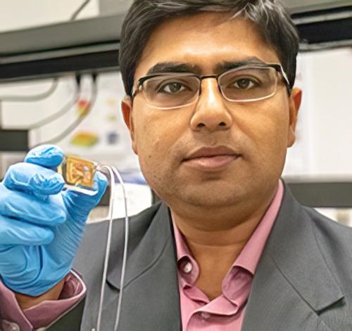
Debashis Chanda, PhD (above), holds up the nanotechnology biosensor he and his team at the University of Central Florida developed that can detect viruses in a blood sample in seconds with 95% accuracy and without the need for pre-preparation of the blood sample. Chanda is professor of physics at the NanoScience Technology Center and the College of Optics and Photonics (CREOL) at UCF. Should this detection device prove effective at instantly detecting viruses at the point of care, clinical laboratories worldwide could have a major new tool in the fight against not just COVID-19, but all viral pathogens. (Photo copyright: University of Central Florida.)
Genetic Virus Detection on a Chip
“The sensitive optical sensor, along with the rapid fabrication approach used in this work, promises the translation of this promising technology to any virus detection, including COVID-19 and its mutations, with high degree of specificity and accuracy,” Debashis Chanda, PhD, told UCF Today. Chanda is professor of physics at the NanoScience Technology Center at UCF and one of the authors of the study. “Here, we demonstrated a credible technique which combines PCR-like genetic coding and optics on a chip for accurate virus detection directly from blood.”
The team tested their device using samples of the Dengue virus that causes Dengue fever, a tropical disease spread by mosquitoes. The device can detect viruses directly from blood samples without the need for sample preparation or purification. This feature enables the testing to be timely and precise, which is critical for early detection and treatment of viruses. The chip’s capability also can help reduce the spread of viruses.
No Pre-processing or Sample Preparation Needed for Multi-virus Testing
The scientists confirmed their device’s effectiveness with multiple tests using varying virus concentration levels and solution environments, including environments with the presence of non-target virus biomarkers.
“A vast majority of biosensors demonstrations in the literature utilize buffer solutions as the test matrix to contain the target analyte,” Chanda told UCF Today. “However, these approaches are not practical in real-life applications because complex biological fluids, such as blood, containing the target biomarkers are the main source for sensing and at the same time the main source of protein fouling leading to sensor failure.”
The researchers believe their device can be easily adapted to detect other viruses and are optimistic about the future of the technology.
“Although there have been previous optical biosensing demonstrations in human serum, they still require off-line complex and dedicated sample preparation performed by skilled personnel—a commodity not available in typical point-of-care applications,” said Abraham Vazquez-Guardado, PhD, a Postdoctoral Fellow at Northwestern University who worked on the study, in the UCS Today article. “This work demonstrated for the first time an integrated device which separated plasma from the blood and detects the target virus without any pre-processing with potential for near future practical usages.”
More research and additional studies are needed to develop the University of Central Florida scientists’ technology and prove its efficacy. However, should the new chip prove viable for point-of-care testing, it would give clinical laboratories and microbiologists an ability to test blood samples without any advanced preparation. Combined with the claims for the device’s remarkable accuracy, that could be a boon not only for COVID-19 testing, but for testing other types of viruses as well.
Researchers say their method can trace ancestry back 100,000 years and could lay groundwork for identifying new genetic markers for diseases that could be used in clinical laboratory tests
Cheaper, faster, and more accurate genomic sequencing technologies are deepening scientific knowledge of the human genome. Now, UK researchers at the University of Oxford have used this genomic data to create the largest-ever human family tree, enabling individuals to trace their ancestry back 100,000 years. And, they say, it could lead to new methods for predicting disease.
This new database also will enable genealogists and medical laboratory scientists to track when, where, and in what populations specific genetic mutations emerged that may be involved in different diseases and health conditions.
New Genetic Markers That Could Be Used for Clinical Laboratory Testing
As this happens, it may be possible to identify new diagnostic biomarkers and genetic indicators associated with specific health conditions that could be incorporated into clinical laboratory tests and precision medicine treatments for chronic diseases.
“We have basically built a huge family tree—a genealogy for all of humanity—that models as exactly as we can the history that generated all the genetic variation we find in humans today,” said Yan Wong, DPhil, an evolutionary geneticist at the Big Data Institute (BDI) at the University of Oxford, in a news release. “This genealogy allows us to see how every person’s genetic sequence relates to every other, along all the points of the genome.”
Researchers from University of Oxford’s BDI in London, in collaboration with scientists from the Broad Institute of MIT and Harvard; Harvard University, and University of Vienna, Austria, developed algorithms for combining different databases and scaling to accommodate millions of gene sequences from both ancient and modern genomes.
“Essentially, we are reconstructing the genomes of our ancestors and using them to form a series of linked evolutionary trees that we call a ‘tree sequence,’” said geneticist Anthony Wilder Wohns, PhD (above), in the Oxford news release. Wohns, a postdoctoral researcher in statistical and population genetics at the Broad Institute, led the study. “We can then estimate when and where these ancestors lived. The power of our approach is that it makes very few assumptions about the underlying data and can also include both modern and ancient DNA samples.” The study may result in new genetic biomarkers that lead to advances in clinical laboratory diagnostics for today’s diseases. (Photo copyright: Harvard School of Engineering and Applied Sciences.)
Tracking Genetic Markers of Disease
The BDI team overcame the major obstacle to tracing the origins of human genetic diversity when they developed algorithms to handle the massive amount of data created when combining genome sequences from many different databases. In total, they compiled the genomic sequences of 3,601 modern and eight high-coverage ancient people from 215 populations in eight datasets.
The ancient genomes included three Neanderthal genomes, a Denisovan genome, and a family of four people who lived in Siberia around 4,600 years ago.
The University of Oxford researchers noted in their news release that their method could be scaled to “accommodate millions of genome sequences.”
“This structure is a lossless and compact representation of 27 million ancestral haplotype fragments and 231 million ancestral lineages linking genomes from these datasets back in time. The tree sequence also benefits from the use of an additional 3,589 ancient samples compiled from more than 100 publications to constrain and date relationships,” the researchers wrote in their published study.
Wong believes his research team has laid the groundwork for the next generation of DNA sequencing.
“As the quality of genome sequences from modern and ancient DNA samples improves, the tree will become even more accurate and we will eventually be able to generate a single, unified map that explains the descent of all the human genetic variation we see today,” he said in the news release.
Developing New Clinical Laboratory Biomarkers for Modern Diagnostics
In a video illustrating the study’s findings, evolutionary geneticist Yan Wong, DPhil, a member of the BDI team, said, “If you wanted to know why some people have some sort of medical conditions, or are more predisposed to heart attacks or, for example, are more susceptible to coronavirus, then there’s a huge amount of that described by their ancestry because they’ve inherited their DNA from other people.”
Wohns agrees that the significance of their tree-recording methods extends beyond simply a better understanding of human evolution.
“[This study] could be particularly beneficial in medical genetics, in separating out true associations between genetic regions and diseases from spurious connections arising from our shared ancestral history,” he said.
The underlying methods developed by Wohns’ team could have widespread applications in medical research and lay the groundwork for identifying genetic predictors of disease risk, including future pandemics.
Clinical laboratory scientists will also note that those genetic indicators may become new biomarkers for clinical laboratory diagnostics for all sorts of diseases currently plaguing mankind.