Proof of vaccination, masking, and availability of on-site testing will continue to be measures taken at in-person events for pathologists and medical laboratory professionals
Organizers of in-person clinical laboratory conferences face an interesting dilemma as they plan events in 2022: Where do they draw the line with COVID-19 safety protocols?
On one hand, the surge of cases caused by the SARS-CoV-2 Omicron variant seems to be in its waning stages and large swaths of the population are vaccinated. On the other hand, clinical laboratory and anatomic pathology events want potential registrants to have confidence that it is safe to travel and attend the gatherings.
One lab industry conference producer who happens to be knee-deep in preparing for an in-person meeting this spring is Robert Michel, Editor-in-Chief of The Dark Report and Founder of the 27th Annual Executive War College on Laboratory and Pathology Management. This informative event takes place on April 27-28 in New Orleans and includes COVID-19 protocols to protect attendees.
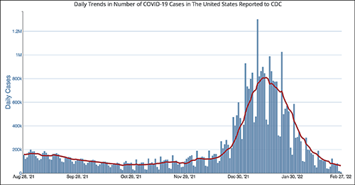
The CDC chart above shows the daily number of new COVID-19 cases in the US for the six-month period ending Feb. 28, 2022. Clinical laboratory managers should note that the number of new cases is at its lowest level since the Omicron variant showed up early this year.
“It’s important for all those planning to attend this year’s Executive War College to know that screening COVID-19 protocols will be in place to ensure the health and safety of all participants,” Michel noted. “We did a large lab conference in the fall of 2021 that included protocols for COVID-19 and the attendees told us they appreciated the protection provided by those protocols.”
After a significant rise in COVID-19 cases in January 2022 due to the Omicron variant, current daily case levels now are lower than they were six months ago before the new variant hit, according to numbers from the federal Centers for Disease Control and Prevention (CDC).
The in-person 2021 Executive War College, which took place in San Antonio on Nov. 2-3, 2021, followed the CDC’s recommendations:
COVID-19 protocols included a daily set of questions and a temperature check for all speakers and attendees before they were allowed to enter the conference area.
CLIA-complex rapid PCR COVID-19 tests were available for individuals whose temperature and answers to the screening questions indicated the need for such testing.
Attendees used an app to answer the daily screening questions and upload proof of vaccination.
“At last fall’s Executive War College, approximately 400 attendees were screened on each of the three days before entering the conference area and not one rapid COVID-19 test was needed,” Michel said. “Not only is that an outstanding outcome, but a number of attendees also told us they appreciated our efforts to keep them safe and protect their health.”
The 2022 Executive War College will follow the CDC’s updated COVID-19 guidelines, along with any state and local directives in effect as of April 27.
Although 300 attendees were expected at the 2021 Executive War College, 400 registered and participated.
Proof of Vaccination Has Been Required at Other Clinical Lab Industry Events
Organizers of other clinical lab conferences also have dealt with COVID-19 safety protocols. For example, the American Clinical Laboratory Association (ACLA) will hold its annual meeting in Washington, D.C., on March 9. COVID-19-related requirements for attendees will include proof of vaccination uploaded to a vaccine verification vendor and proof of a negative PCR test taken within 72 hours prior to the event.
The annual meeting of the American Society of Clinical Pathology (ASCP) occurs later this year in September in Chicago—too early yet to publish protocols. Last year’s ASCP conference in Boston was a hybrid event, offering both in-person and virtual options. Those who attended in person needed to upload proof of vaccination to a third-party vendor and were required to wear masks. On-site COVID-19 testing was available.
Revived Corporate Travel Could Boost Clinical Laboratory Conferences
The path back to live events across all industries has not been easy given various COVID-19 surges, political divisiveness over masking, frozen corporate travel budgets, and corporate policies banning or limiting employee travel.
Conference organizers throughout the United States universally hope those barriers will lower as 2022 progresses.
“With the fast-spreading Omicron triggering another round of setbacks to start 2022, event planners now are betting on spring to finally mark a turning point for the hard-hit industry,” MarketWatch reported on Feb. 4. “Their hopes hinge on American corporations taking a note from the recovery already under way for domestic air travel for leisure purposes, with the linchpin being a robust revival of trade show attendance and other in-person business gatherings.”
For Michel, offering actionable advice through well-thought-out sessions has been a cornerstone of the content offered each year at the Executive War College. He believes that approach will continue to be the strongest drawing point for clinical laboratory and pathology executives now considering attending the event.
“Our reading of the tea leaves is that across the profession of laboratory medicine, a great many managers, administrators, executives, and pathologists want to return to in-person conferences,” Michel noted. “Registrations for our April event are running ahead of 2019, and people tell us that they recognize the changes in healthcare and the lab marketplace because of the pandemic. They want to understand what’s driving current trends, like greater consumer involvement in lab testing and how to get private payers to reimburse claims for COVID-19 and genetic tests, as well as how a growing number of clinical laboratories are incorporating artificial intelligence solutions in both clinical care settings and lab operations.”
International research team that developed swarm learning believe it could ‘significantly promote and accelerate collaboration and information exchange in research, especially in the field of medicine’
“Swarm Learning” is a technology that enables cross-site analysis of population health data while maintaining patient privacy protocols to generate improvements in precision medicine. That’s the goal described by an international team of scientists who used this approach to develop artificial intelligence (AI) algorithms that seek out and identify lung disease, blood cancer, and COVID-19 data stored in disparate databases.
Since 80% of patient records feature clinical laboratory test results, there’s no doubt this protected health information (PHI) would be curated by the swarm learning algorithms.
In their study they wrote, “Fast and reliable detection of patients with severe and heterogeneous illnesses is a major goal of precision medicine. … However, there is an increasing divide between what is technically possible and what is allowed, because of privacy legislation. Here, to facilitate the integration of any medical data from any data owner worldwide without violating privacy laws, we introduce Swarm Learning—a decentralized machine-learning approach that unites edge computing, blockchain-based peer-to-peer networking, and coordination while maintaining confidentiality without the need for a central coordinator, thereby going beyond federated learning.”
What is Swarm Learning?
Swarm Learning is a way to collaborate and share medical research toward a goal of advancing precision medicine, the researchers stated.
The technology blends AI with blockchain-based peer-to-peer networking to create information exchange across a network, the DZNE news release explained. The machine learning algorithms are “trained” to detect data patterns “and recognize the learned patterns in other data as well,” the news release noted.
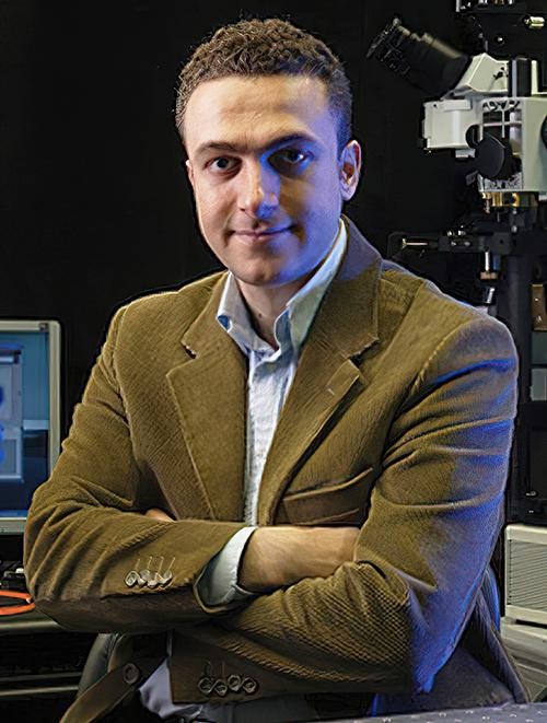
“Medical research data are a treasure. They can play a decisive role in developing personalized therapies that are tailored to each individual more precisely than conventional treatments,” said Joachim Schultze, MD (above), Director, Systems Medicine at DZNE and Professor, Life and Medical Sciences Institute at the University of Bonn, in the news release. “It’s critical for science to be able to use such data as comprehensively and from as many sources as possible,” he added. This, of course, would include clinical laboratory test results data. (Photo copyright: University of Bonn.)
Since, as Dark Daily has reported many times, clinical laboratory test data comprises as much as 80% of patients’ medical records, such a treasure trove of information will most likely include medical laboratory test data as well as reports on patient diagnoses, demographics, and medical history. Swarm learning incorporating laboratory test results may inform medical researchers in their population health analyses.
“The key is that all participants can learn from each other without the need of sharing confidential information,” said Eng Lim Goh, PhD, Senior Vice President and Chief Technology Officer for AI at Hewlett Packard Enterprise (HPE), which developed base technology for swarm learning, according to the news release.
An HPE blog post notes that “Using swarm learning, the hospital can combine its data with that of hospitals serving different demographics in other regions and then use a private blockchain to learn from a global average, or parameter, of results—without sharing actual patient information.
“Under this model,” the blog continues, “‘each hospital is able to predict, with accuracy and with reduced bias, as though [it has] collected all the patient data globally in one place and learned from it,’ Goh says.”
Swarm Learning Applied in Study
The researchers studied four infectious and non-infectious diseases:
They used 16,400 transcriptomes from 127 clinical studies and assessed 95,000 X-ray images.
Data for transcriptomes were distributed over three to 32 blockchain nodes and across three nodes for X-rays.
The researchers “fed their algorithms with subsets of the respective data set” (such as those coming from people with disease versus healthy individuals), the news release noted.
Findings included:
90% algorithm accuracy in reporting on healthy people versus those diagnosed with diseases for transcriptomes.
76% to 86% algorithm accuracy in reporting of X-ray data.
Methodology worked best for leukemia.
Accuracy also was “very high” for tuberculosis and COVID-19.
X-ray data accuracy rate was lower, researchers said, due to less available data or image quality.
“Our study thus proves that swarm learning can be successfully applied to very different data. In principle, this applies to any type of information for which pattern recognition by means of artificial intelligence is useful. Be it genome data, X-ray images, data from brain imaging, or other complex data,” Schultze said in the DZNE news release.
The scientists say hospitals as well as research institutions may join or form swarms. So, hospital-based medical laboratory leaders and pathology groups may have an opportunity to contribute to swarm learning. According to Schultze, sharing information can go a long way toward “making the wealth of experience in medicine more accessible worldwide.”
Though the new technology could speed diagnoses of cancers and other skin diseases, it would also greatly reduce dermatopathology biopsy referrals and revenue
What effect would elimination of tissue biopsies have on dermatopathology and clinical laboratory revenue? Quite a lot. Dermatologists alone account for a significant portion of skin biopsies sent to dermatopathologists. Thus, any new technology that can “eliminate the need for invasive skin biopsies” would greatly reduce the number of histopathological referrals and reduce revenue to those practices.
“What if we could entirely bypass the biopsy process and perform histology-quality staining without taking tissue and processing tissue in a noninvasive way? Can we create images that diagnosticians can benefit from?” asked Aydogan Ozcan, PhD (above), Chancellor’s Professor of Electrical and Computer Engineering at UCLA’s Samueli School of Engineering, one of the scientists who developed UCLA’s new virtual histology method, during an interview with Medical Device + Diagnostic Industry (MD+DI). (Photo copyright: Nature.)
Could Skin Biopsies be Eliminated?
The UCLA researchers believe their innovative deep learning-enabled imaging framework could possibly circumvent the need for skin biopsies to diagnose skin conditions.
“Here, we present a deep learning-based framework that uses a convolutional neural network to rapidly transform in vivo RCM images of unstained skin into virtually-stained hematoxylin and eosin-like images with microscopic resolution, enabling visualization of the epidermis, dermal-epidermal junction, and superficial dermis layers.
“This application of deep learning-based virtual staining to noninvasive imaging technologies may permit more rapid diagnoses of malignant skin neoplasms and reduce invasive skin biopsies,” the researchers added in their published study.
According to the published study, the UCLA team trained their neural network under an adversarial machine learning scheme to transform grayscale RCM images into virtually stained 3D microscopic images of normal skin, basal cell carcinoma, and pigmented melanocytic nevi. The new images displayed similar morphological features to those shown with the widely used hematoxylin and eosin (H&E) staining method.
“In our studies, the virtually stained images showed similar color contrast and spatial features found in traditionally stained microscopic images of biopsied tissue,” Ozcan told Photonics Media. “This approach may allow diagnosticians to see the overall histological features of intact skin without invasive skin biopsies or the time-consuming work of chemical processing and labeling of tissue.”
The framework covers different skin layers, including the epidermis, dermal-epidermis, and superficial dermis layers. It images deeper into tissue without being invasive and can be quickly performed.
“The virtual stain technology can be streamlined to be almost semi real time,” Ozcan told Medical Device + Diagnostic Industry (MD+DI). “You can have the virtual staining ready when the patient is wrapping up. Basically, it can be within a couple of minutes after you’re done with the entire imaging.”
Currently, medical professionals rely on invasive skin biopsies and histopathological evaluations to diagnose skin diseases and cancers. These diagnostic techniques can result in unnecessary biopsies, scarring, multiple patient visits and increased medical costs for patients, insurers, and the healthcare system.
Improving Time to Diagnosis through Digital Pathology
Another advantage of this virtual technology, the UCLA researchers claim, is that it can provide better images than traditional staining methods, which could improve the ability to diagnose pathological skin conditions and help alleviate human error.
“The majority of the time, small laboratories have a lot of problems with consistency because they don’t use the best equipment to cut, process, and stain tissue,” dermatopathologist Philip Scumpia, MD, PhD, Assistant Professor of Dermatology and Dermatopathology at UCLA Health and one of the authors of the research paper, told MD+DI.
“What ends up happening is we get tissue on a histology slide that’s basically unevenly stained, unevenly put on the microscope, and it gets distorted,” he added, noting that this makes it very hard to make a diagnosis.
Scumpia also added that this new technology would allow digital images to be sent directly to the pathologist, which could reduce processing and laboratory times.
“With electronic medical records now and the ability to do digital photography and digital mole mapping, where you can obtain a whole-body imaging of patients, you could imagine you can also use one of these reflectance confocal devices. And you can take that image from there, add it to the EMR with the virtual histology stain, which will make the images more useful,” Scumpia said. “So now, you can track lesions as they develop.
“What’s really exciting too, is that there’s the potential to combine it with other artificial intelligence, other machine learning techniques that can give more information,” Scumpia added. “Using the reflectance confocal microscope, a clinician who might not be as familiar in dermatopathology could take images and send [them] to a practitioner who could give a more expert diagnosis.”
Faster Diagnoses but Reduced Revenue for Dermatopathologists, Clinical Labs
Ozcan noted that there’s still a lot of work to be done in the clinical assessment, validation, and blind testing of their AI-based staining method. But he hopes the technology can be propelled into a useful tool for clinicians.
“I think this is a proof-of-concept work, and we’re very excited to make it move forward with further advances in technology, in the ways that we acquire 3D information [and] train our neural networks for better and faster virtual staining output,” he told MD+DI.
Though this new technology may reduce the need for invasive biopsies and expedite the diagnosis of skin conditions and cancers—thus improving patient outcomes—what affect might it have on dermatopathology practices?
More research and clinical studies are needed before this new technology becomes part of the diagnosis and treatment processes for skin conditions. Nevertheless, should virtual histology become popular and viable, it could greatly impact the amount of skin biopsy referrals to pathologists, dermatopathologists, and clinical laboratories, thus diminishing a great portion of their revenue.
Japanese scientists who developed the detection method hope to use it to create ‘easy testing kits that anyone can use’
What do ostriches and humans have in common during the current COVID-19 pandemic? The unexpected answer is that ostrich antibodies can be used to identify humans infected with COVID-19. If proven viable in healthcare settings, the possibility exists that new clinical laboratory tests could be developed based on wearable diagnostics technologies that pathologists would interpret for doctors and patients.
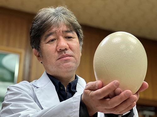
According to Study Finds, scientists at Kyoto Prefectural University in Japan have created a removable mask filter that, when sprayed with a fluorescent dye coated with antibodies extracted from ostrich eggs, will glow under UV light when COVID-19 is detected. The discovery by Yasuhiro Tsukamoto, PhD (above), President of Kyoto Prefectural University, and his researchers could lead to development of low-cost at home COVID-19 testing kits using the same ostrich-antibody-based technique. (Photo copyright: Kyoto Prefectural University/Reuters.)
The KPU scientists conducted a small study with 32 COVID-19 patients over a 10-day span. The surgical-style masks they wore later glowed around the nose and mouth areas but became dimmer over time as their viral load decreased.
“The ostrich antibody for corona placed on the mouth filter of the mask captures the coronavirus in coughing, sneezing, and water,” the researchers explained in Study Finds.
Tsukamoto himself learned he had contracted COVID-19 after wearing a prototype mask and noticing it glowed under UV light. A PCR test later confirmed his diagnosis, Kyodo News reported.
The KPU team “hopes to further develop the masks so they will glow automatically, without special lighting, if the [COVID-19] virus is detected.” Reuters noted in its coverage of the ostrich-antibody masks.
Making Medicine from Ostrich Antibodies
A profile in Audubon noted that Tsukamoto, who also serves as a veterinary medicine professor at Kyoto Prefectural University, made ostriches the focus of his research since the 1990s as he looked for ways to harness the dinosaur-like bird’s properties to fight human infections. He maintains a flock of 500 captive ostriches. Each female ostrich can produce 50 to 100 eggs/year over a 50-year life span.
Tsukamoto’s research focuses on customizing the antibodies in ostrich eggs by injecting females with inactive viruses, allergens, and bacteria, and then extracting the antibodies to develop medicines for humans. Antibodies form in the egg yolks in about six weeks and can be collected without harming the parent or young.
“The idea of using ostrich antibodies for therapeutics in general is a very interesting concept, particularly because of the advantages of producing the antibodies from eggs,” Ashley St. John, PhD, an Associate Professor in Immunology, at Duke-NUS Medical School in Singapore, told Audubon.
While more clinical studies will be needed before ostrich-antibody masks reach the commercial marketplace, Tsukamoto’s team is planning to expand their experiment to 150 participants with a goal of receiving Japanese government approval to begin selling the glowing COVID-detection masks as early as 2022. But they believe the ostrich-antibody technique ultimately may lead to development of an inexpensive COVID-19 testing kit.
“We can mass-produce antibodies from ostriches at a low cost. In the future, I want to make this into an easy testing kit that anyone can use,” Tsukamoto told Kyodo News.
Harvard, MIT Also Working on COVID-19 Detecting Facemask
According to Fast Company, the MIT/Harvard COVID-19-detecting masks use the same core technology as previous paper tests for Ebola and Zika that utilize proteins and nucleic acids embedded in paper that react to target molecules.
Fast Company explained that the mask wearer launches a test by pushing a button to release a small water reservoir embedded in the mask (above). Droplets from their breath are than analyzed by the sensors in the masks, which could be adapted to test for new COVID variants or other respiratory pathogens. In addition to eliminating the use of a nasal swab, the mask-based testing system may compete with clinical laboratory-based results. (Photo copyright: Felice Frankel/MIT.)
“They would especially be useful in situations where local variant outbreaks are occurring, allowing people to conveniently test themselves at home multiple times a day,” he told Fast Company.
“It’s on par specificity and sensitivity that you will get in a state-of-the-art [medical] laboratory, but with no one there,” Luis Ruben Soenksen, PhD, Venture Builder in Artificial Intelligence and Healthcare at MIT and one of the co-authors of the Nature Biotechnology study, told Fast Company.
As the definition of “wearable diagnostic technology” broadens, pathologists and clinical laboratory scientists may see their roles expand to include helping consumers interpret data collected by point-of-care testing technology as well as performing, evaluating, and interpreting laboratory test results that come from non-traditional sources.
GI pathologists will be interested in how the Endoculus device uses tank-like treads to traverse the gastrointestinal tract, where it can capture images and perform biopsies
Gastroenterologists (GI) may soon gain a useful new tool for use in gathering both biopsies and diagnostic information when examining the gastrointestinal tract. Ongoing development of a new robotic device promises both capabilities using technology that will be of interest to GI pathologists and clinical laboratory scientists.
The minute robotic device uses tank-like treads to traverse the colon. While there, it can capture live images and perform biopsies under the control of a gastroenterologist. The researchers believe the robotic technology will benefit GIs performing the colonoscopies as well as the pathologists called upon to analyze biopsies.
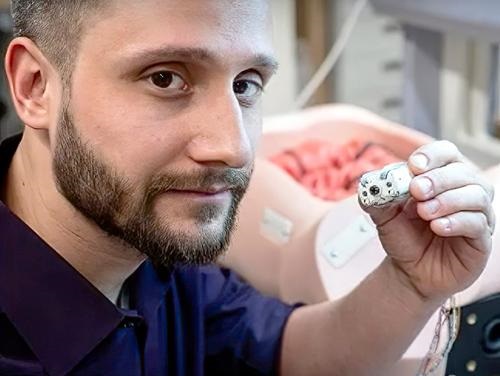
“Currently, endoscopy consists of a gastroenterologist using a semi-rigid, long rope-like device and endoscope to propel through your colon manually,” Gregory Formosa, PhD (above) a member of the AMTL team that developed Endoculus, said in a YouTube video describing the device. “We think that a robotic capsule endoscope can replace conventional endoscopes by making them faster, safer, and more robust than a human operator can do currently with traditional techniques,” he added. (Photo copyright: University of Colorado.)
AMTL researcher Gregory Formosa, PhD, said the team’s goal is to “have a capsule-sized robot that can actively traverse [a patient’s] entire gastrointestinal tract and send out diagnostics in real time, as well as autonomously navigate itself to localize problematic areas within [the] intestinal tract.”
Formosa noted that colorectal cancer is “the third-most fatal and diagnosed cancer in the United States.” But if caught at an early stage, these cancers are “95% treatable,” he added. “So, if we can get people screened early, we definitely can reduce the fatality rate of colorectal cancers significantly.”
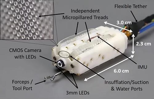
Currently about the size of a C battery, Endoculus (above) is a “fully packed medical device, complete with a camera, an air pump for inflating the colon, a water pump for cleaning, and a tool port for holding biopsy snares,” states a University of Colorado news story, titled, “A Robot May One Day Perform Your Colonoscopy.” (Photo copyright: University of Colorado.)
How Endoculus Works
One key to the device are the four treads, which are designed for traction on digestive tissue.
“You have to forget about everything you know from a locomotion standpoint because driving around inside the body is very different than driving around in a car,” said Rentschler in the University of Colorado news story. “The environment is highly deformable. It’s very slick. There are sharp peaks that you have to go over.”
The university news story noted the current availability of ingestible “pill cams” that can take photos as they travel through the digestive system. But once swallowed, their movements cannot be controlled.
“For our robots to be able to reach those regions that [can be] reached with a pill-cam—but also be able to stop and look around—that could be a big paradigm shift in the way we view these procedures,” said Micah Prendergast, PhD, an AMTL research team member.
Could Biopsies Be Diagnosed In Situ with Endoculus?
The researchers currently view Endoculus as a potentially better way to perform conventional biopsies. But could it lead to bigger advancements?
“Researchers continue to develop devices to help various specialist physicians—in this case GIs—do more when treating patients,” said Dark Daily Publisher and Editor-in-Chief Robert Michel. “This device fits that description. It is designed to improve the ability of GIs to evaluate the colon. Not only does this device do that, but it can also collect a biopsy at sites of interest. In this way, it is a device that can be a benefit to pathologists who will analyze the biopsy.
“With improvements in digital cameras and associated AI-powered analytical tools, the day might not be far off when a device like this can use the camera and artificial intelligence to diagnose the tissue of interest in situ,” he added. “This might create the opportunity for pathologists to be present in the exam room during the procedure, or even viewing the images remotely.
“Not only would that eliminate the need to collect a tissue specimen that must then be sent to a pathology lab, but it would create a new opportunity for pathologists to add value to patient care while shortening the time to diagnosis for the tissue of interest during these procedures,” Michel noted.