Newly combined digital pathology, artificial intelligence (AI), and omics technologies are providing anatomic pathologists and medical laboratory scientists with powerful diagnostic tools
Add “spatial transcriptomics” to the growing list of “omics” that have the potential to deliver biomarkers which can be used for earlier and more accurate diagnoses of diseases and health conditions. As with other types of omics, spatial transcriptomics might be a new tool for surgical pathologists once further studies support its use in clinical care.
Among this spectrum of omics is spatial transcriptomics, or ST for short.
Spatial Transcriptomics is a groundbreaking and powerful molecular profiling method used to measure all gene activity within a tissue sample. The technology is already leading to discoveries that are helping researchers gain valuable information about neurological diseases and breast cancer.
Marriage of Genetic Imaging and Sequencing
Spatial transcriptomics is a term used to describe a variety of methods designed to assign cell types that have been isolated and identified by messenger RNA (mRNA), to their locations in a histological section. The technology can determine subcellular localization of mRNA molecules and can quantify gene expression within anatomic pathology samples.
In “Spatial: The Next Omics Frontier,” Genetic Engineering and Biotechnology News (GEN) wrote, “Spatial transcriptomics gives a rich, spatial context to gene expression. By marrying imaging and sequencing, spatial transcriptomics can map where particular transcripts exist on the tissue, indicating where particular genes are expressed.”
In an interview with Technology Networks, George Emanuel, PhD, co-founder of life-science genomics company Vizgen, said, “Spatial transcriptomic profiling provides the genomic information of single cells as they are intricately spatially organized within their native tissue environment.
“With techniques such as single-cell sequencing, researchers can learn about cell type composition; however, these techniques isolate individual cells in droplets and do not preserve the tissue structure that is a fundamental component of every biological organism,” he added.
“Direct spatial profiling the cellular composition of the tissue allows you to better understand why certain cell types are observed there and how variations in cell state might be a consequence of the unique microenvironment within the tissue,” he continued. “In this way, spatial transcriptomics allows us to measure the complexity of biological systems along the axes that are most relevant to their function.”
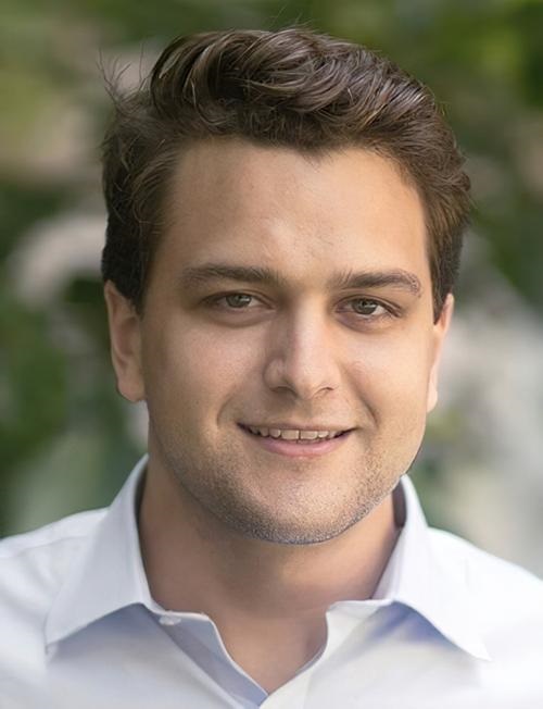
“Although spatial genomics is a nascent field, we are already seeing broad interest among the community and excitement across a range of questions, all the way from plant biology to improving our understanding of the complex interactions of the tumor microenvironment,” George Emanuel, PhD (above), told Technology Networks. Oncologists, anatomic pathologists, and medical laboratory scientists my soon see diagnostics that take advantage of spatial genomics technologies. (Photo copyright: Vizgen.)
According to 10x Genomics, “spatial transcriptomics utilizes spotted arrays of specialized mRNA-capturing probes on the surface of glass slides. Each spot contains capture probes with a spatial barcode unique to that spot.
“When tissue is attached to the slide, the capture probes bind RNA from the adjacent point in the tissue. A reverse transcription reaction, while the tissue is still in place, generates a cDNA [complementary DNA] library that incorporates the spatial barcodes and preserves spatial information.
“Each spot contains approximately 200 million capture probes and all of the probes in an individual spot share a barcode that is specific to that spot.”
“The highly multiplexed transcriptomic readout reveals the complexity that arises from the very large number of genes in the genome, while high spatial resolution captures the exact locations where each transcript is being expressed,” Emanuel told Technology Networks.
Spatial Transcriptomics for Breast Cancer and Neurological Diagnostics
In that paper, the authors wrote “we envision that in the coming years we will see simplification, further standardization, and reduced pricing for the ST protocol leading to extensive ST sequencing of samples of various cancer types.”
Spatial transcriptomics is also being used to research neurological conditions and neurodegenerative diseases. ST has been proven as an effective tool to hunt for marker genes for these conditions as well as help medical professionals study drug therapies for the brain.
“You can actually map out where the target is in the brain, for example, and not only the approximate location inside the organ, but also in what type of cells,” Malte Kühnemund, PhD, Director of Research and Development at 10x Genomics, told Labiotech.eu. “You actually now know what type of cells you are targeting. That’s completely new information for them and it might help them to understand side effects and so on.”
The field of spatial transcriptomics is rapidly moving and changing as it branches out into more areas of healthcare. New discoveries within ST methodologies are making it possible to combine it with other technologies, such as Artificial Intelligence (AI), which could lead to powerful new ways oncologists and anatomic pathologists diagnose disease.
“I think it’s going to be tricky for pathologists to look at that data,” Kühnemund said. “I think this will go hand in hand with the digital pathology revolution where computers are doing the analysis and they spit out an answer. That’s a lot more precise than what any doctor could possibly do.”
Spatial transcriptomics certainly is a new and innovative way to look at tissue biology. However, the technology is still in its early stages and more research is needed to validate its development and results.
Nevertheless, this is an opportunity for companies developing artificial intelligence tools for analyzing digital pathology images to investigate how their AI technologies might be used with spatial transcriptomics to give anatomic pathologists a new and useful diagnostic tool.
Self-insured and campus health markets are contract opportunities for small and midsize clinical laboratories through investment in data infrastructure and management
Bordenave spoke this week at the Executive War College in San Antonio. During two intriguing presentations, she shared that the self-insured employer and campus health markets are areas of opportunity for small and midsize clinical laboratories. This is because employer groups and college campuses are busy communities of covered individuals, and these population health groups are well-suited for proactive care models.
In fact, she said, some clinical laboratories may already be well-positioned to serve these customers.
Self-Insured Employer Groups and Campus Health Markets as New Clinical Laboratory Customers
According to CMS national health expenditure data, in 2020, a whopping $4 trillion was spent on healthcare in the US. In the middle of all that are people living, going to school, and working who have high blood pressure, rising lipid levels, lower-back pain, migraines, and other health conditions waiting to be diagnosed and flagged for follow-up.
And as pathologists and clinical laboratory managers know, 80% of those healthcare encounters result in lab test data.
Clinical laboratories, therefore, can gain customers among self-insured employer groups and similarly functioning campus health markets that serve students.
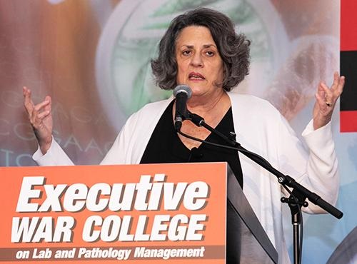
During her presentations at the 2021 Executive War College in San Antonio, Kristine Bordenave, MD, FACP (above), a strategic consultant in precision medicine, population health, Medicare compliance, and cost management, noted that “just about all paths forward post-COVID will require the data infrastructure of clinical laboratories to achieve an advanced level of functionality.” (Photo copyright: The Dark Intelligence Group.)
In one example she gave during her presentation, Bordenave noted that self-insured employer groups “were more than willing to contract directly, and they were contracting for care that directly relates to lab. Anything that would help reduce presenteeism and absenteeism with their employees.”
Presenteeism and Absenteeism
For years, presenteeism and absenteeism have plagued employee productivity in organizations large and small. Both have been attributed to numerous individual health and wellness factors among individuals. At some point, these issues culminate into various forms of reactive healthcare services and safety issues, she added.
The cost of presenteeism is estimated at between $150 billion and $225 billion. Meanwhile, at least 60% of employees are now covered in fully-funded or partially-funded self-insured plans, Healthcare Finance reported.
The way a campus health system operates is similar to a self-insured model but more of an integrated delivery system, Bordenave said. Among the priorities are controlling the spread of infectious diseases, such as COVID-19 and measles.
Clinical Laboratory Data Valuable in Treating-to-Goal and Closing Care Gaps
During two featured Executive War College general session discussions, Bordenave explained the focus of her work: aligning primary care with the clinical laboratory to treat-to-goal and close care gaps.
“There was a lot of focus on us taking laboratory information and treating people to goal, and that was with respect to diabetes, cholesterol, and hypertension, because those are three common diseases that exist within their [employee] populations. [Primary care doctors] know [that] if they [can] maximize the care in those patients—so that the patient is maximally treated—that patient performs. There’s a lot of literature around this.”
In the state of New Mexico where Bordenave’s project evolved, a culture of innovation prevails, where like-minded people have an opportunity to “do the unique,” she explained. The state’s population is spread out, there is a shortage of healthcare providers, and people generally lack access to health services and other social determinants of health. The liberty to think outside the box—to ensure care in creative ways—was essential to the success of Bordenave’s project.
“Blue Cross Blue Shield paid handsomely for improving healthcare outcomes in diabetes,” she said, adding, “and we never did a standard visit with any of those patients, ever. Then we got paid by a big employer group to do the same thing for them.”
Future of Clinical Laboratory Functionality
Bordenave noted that just about all paths forward post-COVID will require the data infrastructure of clinical laboratories to achieve an advanced level of functionality. Dark Daily will cover more opportunities for labs to capitalize on their structured data in future ebriefings.
Executive War College is scheduled to reconvene April 27-28, 2022, in New Orleans. In the meantime, recordings of this year’s presentations will be available for download, including:
A Roundtable Discussion on Current Activity Involving Clinical Laboratory and Pathology Mergers and Acquisitions.
Taking a Deeper Dive into How Artificial Intelligence Analyzes a Digital Pathology Image: What Current Technology Can and Cannot Do, Steps to Implement, and Understanding How the FDA Views AI in Digital Pathology.
Open Conversation About the Healthcare Data Aggregation Hub Model.
And more.
To learn about Executive War College’s complete program package, send an email request to info@darkreport.com.
One of the world’s fastest growing medical laboratory companies in India is using digital pathology systems and AI to replace older diagnostic technologies
Artificial intelligence (AI) is gaining acceptance around the world and use of AI to analyze digital pathology images is expected to be a major disruptor to the profession of anatomic pathology. Internationally, several pathology companies already use AI-powered solutions to diagnose cancer.
One such example is Neuberg Diagnostics, a fast-growing clinical laboratory company in Chennai, India. Neuberg has been using AI to review digital pathology images for several years, according to Chairman and Managing Director GSK Velu, PhD, BPharm.
“We already use AI in our laboratories,” Velu said in an exclusive interview with Dark Daily. “Our main reference laboratories currently use digital pathology systems to support the pathologists and many of them are using AI with these digital pathology systems.
“AI and data analytics tools are being used in other departments too, such as in our wellness department where we use AI for predictive analytics,” he added. “We also use AI in our genomics division, and we are introducing AI into other divisions slowly and steadily.”
Neuberg operates 120 laboratories in an extensive network in India, South Africa, and the United Arab Emirates (UAE), and now in the US as well.
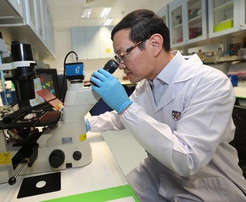
“Our idea is to enhance the access and affordability for next-generation techniques, meaning molecular diagnostics, genomics, pathology, digital pathology, proteomics, metabolomics, and all that. This is the spirit behind Neuberg Diagnostics,” said GSK Velu, PhD, BPharm (above), Chairman and Managing Director of Neuberg Diagnostics, in an exclusive interview with The Dark Report. Clinical laboratories that are considering investing in digital pathology technologies may want to follow its development at Neuberg’s Centre for Genomic Medicine in Raleigh, NC, which opened in May. (Photo copyright: Neuberg Diagnostics.)
Replacing Older Pathology Technologies
As has been happening at other anatomic pathology centers around the world, Neuberg has been using digital pathology systems to replace older technologies. “One of our largest labs is our Bangalore Reference Lab,” Velu said. “There, we do not use microscopes for histopathology, and that lab has used digital pathology for routine review of specimens for several years now.
“But because artificial intelligence is still emerging, we can’t rely on AI with all of our digital pathology systems,” he added. “Although, of course, AI is certainly an aid to everything we do with digital pathology.
“For a variety of reasons, the adaptation of artificial intelligence in anatomic pathology is not happening as effectively nor as fast as we would like,” he noted. “So, for now, we need to wait and watch a bit longer, either because adaptation by pathologists is slow, or because AI tools are still a bit of a worry for some pathologists.
Younger Pathologists Adapt Faster to Digital Pathology
One reason could be that conventional pathologists worry about relying completely on AI for any diagnosis, Velu noted. “I’m certain that the more recent generation of pathologists who are now in their 30s, and the new people coming into pathology, will start adapting more quickly to digital pathology and to AI faster than the older generation of pathologists have done.
“The younger pathologists have a greater appreciation for the potential of digital pathology, while the older pathologists don’t want to let go of conventional diagnosis methods,” he added.
“For example, we have not yet seen where pathologists are reviewing breast image scans,” he commented. “But, at the same time, AI has been well-accepted among radiologists who are reviewing breast mammography scans.”
In India and in other markets worldwide, radiologists have adapted AI tools for breast mammography scans to diagnose breast cancer, he noted. “But that’s not happening even among pathologists who are doing cancer screening,” he said.
Velu suggested that another reason for the slow adoption of AI tools in pathology is that these systems are relatively new to the market. “Maybe the AI tools that are used with digital pathology are not as reliable as we hoped they would be, or they are not fully robust at the moment,” he speculated. “That’s why I say it will take some time before the use of AI for diagnosis becomes more widespread among pathologists. So, for now, we must wait until digital pathology and AI tools work together more seamlessly.
Replacing Conventional Pathology Technologies and Methods
“When those two technologies—AI and digital pathology systems—are linked more closely, their use will take hold in a substantial way,” Velu predicted. “When that happens, they are likely to replace conventional pathology methods completely.
“Currently, we are in the early stages of a transformation,” he added. “In our labs, you can see that the transformation is ongoing. We are using digital pathology systems even in our smaller labs. Then, the staff in our smaller labs do the processing of slides to convert them to digital images and send them to our labs in the larger cities. There, the professional staff uses AI to review those digital images and issue reports based on those images.
“Using our digital pathology systems and AI in that way means that we can make that technology available even in smaller towns and villages that have access only to our smaller labs,” he commented.
Velu added that wider use of digital pathology systems could improve the quality of care that pathologists deliver to patients in a significant way, particularly in rural areas. “Here in India, we are not seeing a huge shortage of pathologists, except in rural areas and villages,” he explained. “In those places, we could run short of pathologists.
“That is the reason we are trying to adapt the use of telepathology more widely,” he noted. “To do that, we might have technicians and histologists who will do just processing of slides so that they can send the digital images to our pathologists located in larger cities. Then, those surgical pathologists will review the cases and send the reports out. That’s the model that we are trying to slowly follow here.”
As use of digital pathology images increased, many predicted that specimens would flow from the US to India. This would happen because of the belief that the lower cost of surgical pathology in India would successfully draw business away from pathology groups here in the United States.
However, Neuberg turned the tables on that belief when it announced the opening of its Neuberg Centre for Genomic Medicine (NCGM), a state-of-the-art esoteric and genetic testing laboratory in Raleigh, NC. The NCGM lab is CLIA-certified and Neuberg says it is ready to compete with labs in this country on their home turf.
These are reasons why pathologists and pathology practice administrators in the United States may want to watch how Neuberg Diagnostics continues to develop its use of digital pathology platforms and AI-powered digital image analysis tools throughout its international network of laboratories.
Webinar: Roundtable Discussion of Digital Pathology Adoption Across Key Functional Areas: Diagnostic Pathology, Education, and Research PRESS RELEASE FOR IMMEDIATE RELEASE THE DARK REPORT21806 Briarcliff Dr.Spicewood, TX 78669512-264-7103 o512-264-0969 f Media Contact: Bill Sinagrainfo@darkreport.com AUSTIN, Texas (September 21, 2021) —To help pathologists and pathology practice administrators adopt and develop a digital pathology strategy, Dark Daily presents a new free webinar at 1pm Eastern...
The new method employs a pH sensitive dye and AI algorithms to ‘distinguish between cells originating from normal and cancerous tissue, as well as among different types of cancer’ the researchers said
Might a pH-sensitive dye in tandem with an image analysis solution soon be used to identify cancerous cells within blood samples as well within tissue? Recent research indicates that could be a possibility. If further studies and clinical trials confirm this capability, then anatomic pathologists could gain another valuable tool to use in diagnosing cancers and other types of disease.
Currently, surgical pathologists use a variety of hematoxylin and eosin stains (H/E) to bring out useful features in cells and cell structures. So, staining tissue on glass slides is a common practice. Now, thanks to machine learning and artificial intelligence, anatomic pathologists may soon have a similar tool for spotting cancer cells within both tissue and blood samples.
Researchers at the National University of Singapore (NUS) have developed a method for identifying cancer that uses a pH sensitive dye called bromothymol blue. The dye reacts to various levels of acidity in cancer cells by turning colors. “The pH inside cancer cells tends to be higher than that of healthy cells. This phenomenon occurs at the very early phases of cancer development and becomes amplified as it progresses,” Labroots reported.
In “Machine Learning Based Approach to pH Imaging and Classification of Single Cancer Cells,” published in the journal APL Bioengineering, the NUS researchers wrote, “Here, we leverage a recently developed pH imaging modality and machine learning-based single-cell segmentation and classification to identify different cancer cell lines based on their characteristic intracellular pH. This simple method opens up the potential to perform rapid noninvasive identification of living cancer cells for early cancer diagnosis and further downstream analyses.”
According to an NUS news release, the bromothymol blue dye is “applied onto patients’ cells” being held ex vivo in cell culture dishes. The dye’s color changes depending on the acidity level of the cancer cells it encounters. Microscopic images of the now-visible cancers cells are taken, and a machine-learning algorithm analyzes the images before generating a report for the anatomic pathologist.
The NUS researchers claim the test can provide answers in about half an hour with 95% accuracy, Labroots reported.
“The ability to analyze single cells is one of the holy grails of health innovation for precision medicine or personalized therapy. Our proof-of-concept study demonstrates the potential of our technique to be used as a fast, inexpensive and accurate tool for cancer diagnosis,” said Lim Chwee Teck, PhD, NUS Society Professor and Director of NUS’ Institute for Health Innovation and Technology, in the NUS news release.
The novel technique for differentiating cancer cells from non-cancerous cells being developed at the National University of Singapore (NUS) could eventually become useful in detecting cancer cells in tissue samples, either obtained from tumor biopsies or blood samples. “As the number of cells in these samples can be in millions or even billions, the ability to detect the very few cancer cells among the others will be useful for clinicians,” NUS Society Professor and Director of NUS’ Institute for Health Innovation and Technology, Lim Chwee Teck, PhD (above) told The Straits Times. (Photo copyright: The Straits Times.)
AI Cell Analysis versus Laborious Medical Laboratory Steps
By developing an AI-driven method, Professor Lim and the NUS team sought to improve upon time-consuming techniques for identifying cells that traditionally involve using florescent probes, nanoparticles, and labeling steps, or for cells to be fixed or terminated.
“Unlike other cell analysis techniques, our approach uses simple, inexpensive equipment, and does not require lengthy preparation and sophisticated devices. Using AI, we are able to screen cells faster and accurately,” Professor Lim told Labroots. “Furthermore, we can monitor and analyze living cells without causing any toxicity to the cells or the need to kill them.”
The new technique may have implications for cancer detection in tumor tissue as well as in liquid biopsies.
“We are also exploring the possibility of performing the real-time analysis on circulating cancer cells suspended in blood,” Professor Lim said in the NUS news release. “One potential application for this would be in liquid biopsy where tumor cells that escaped from a primary tumor can be isolated in a minimally-invasive fashion from bodily fluids such as blood.”
Diagnosing Cancer in Real Time
The NUS’ method requires more research and clinical studies before it could become an actual tool for anatomic pathologists and other cancer diagnosticians. Additionally, the NUS researchers acknowledged that the focus on only four cell lines (normal cells, benign breast tumor cells, breast cancer cells, and pancreatic cancer cells) limited their study, as did lack of comparison with conventional florescent pH indicators.
Still, the NUS scientists are already planning more studies to advance their concept to different stages of cell malignancy. They envision a “real-time” version of the technique to enable recognition of cells and fast separation of those that need to be referred to clinical laboratories for molecular testing and/or genetic sequencing.
Medical laboratory leaders may want to follow the NUS study. An inexpensive AI-driven method that can accurately detect and classify cancer cells based on pH within the cells is provocative and may be eventually become integrated with other cancer diagnostics.