Faulkner was surpassed on Forbes’ list only by roofing material magnate Diane Hendricks, co-founder of ABC Supply Co., whose net worth of $11 billion puts her squarely in the top spot.
Richest Self-Made Women in Healthcare
Becker’s Hospital Review highlighted the seven richest “self-made” women who ran healthcare-related companies. They include:
Judith Faulkner, founder and CEO of Epic, ranked 2nd, net worth $6.5 billion.
Alice Schwartz, co-founder of Bio-Rad Laboratories, ranked 10th, net worth $2.9 billion.
Heather Hasson and Trina Spear, co-founders and co-CEOs of FIGS (direct-to-consumer healthcare apparel and scrubs), ranked 50th and 52nd, net worth $625 million and $600 million respectively.
Also listed by Forbes was Anne Wojcicki, CEO and founder of 23andMe, a personal genomics and biotechnology company. Wojcicki’s net worth of $1.1 billion puts her in the 25th position, according to Forbes.
In “Genetic Test Company 23andMe Completes Merger with Richard Branson’s VG Acquisition Corp., Stock Now Trades on NASDAQ,” Dark Daily noted that since the Sunnyvale, Calif. direct-to-consumer (DTC) genetic testing company will now be filing quarterly earnings reports, pathologists and clinical laboratory managers will have the opportunity to learn more about how 23andMe serves the consumer market for genetic types and how it is generating revenue from its huge database containing the genetic sequences from millions of people.
Judith Faulkner (left), founder and CEO of Epic Systems Corp., and Alice Schwartz (right), co-founder of Bio-Rad Laboratories, ranked 2nd and 10th respectively in Forbes’ list of the top 100 richest self-made women. In healthcare, Faulkner ranks 1st and Schwartz 2nd. Clinical laboratory personnel will likely be familiar with Epic Beaker, which, according to Healthcare IT Leaders, “is Epic’s laboratory information system (LIS) for hospitals, clinics, patient service centers, and reference labs. The software supports common workflows for clinical pathology (CP) labs as well as anatomic pathology (AP) labs.” (Photo copyrights: HIT Consultant/Science History Institute.)
“I always liked making things out of clay. And the computer was clay of the mind. Instead of physical, it was mental,” Faulkner, who is 77, told Forbes.
Company milestones noted by Forbes include:
Inking a deal in 2004 with Kaiser Permanente for a three-year, $400-million project.
Moving in 2005 to a corporate campus in southern Wisconsin—an “adult Disney World” with the largest underground auditoriums and more “fantastical” buildings.
More recently, AdventHealth of Altamonte Springs, Fla., contracted with Epic for a $650 million remote build and installation.
“Epic’s system has tentacles that go out through amazing networks. You can actually help a person get the care they need wherever they need to get it,” AdventHealth’s CEO Terry Shaw told Forbes.
“I think that what will happen is that a few of them will do very well. And the majority of them won’t. “It’s not us as much as the health systems who have to respond to the patient saying, ‘Send my data here,’ or ‘Send my data there,’” Faulkner told Forbes.
Bio-Rad’s Alice Schwartz an IVD ‘Pioneer’
As Faulkner rose to prominence in healthcare IT, Alice Schwartz of Bio-Rad Laboratories found massive success in the in vitro diagnostics industry.
She and her late husband, David, started Bio-Rad with $720 in 1952 in Berkeley, Calif. They were intent on offering life science products and services aimed at identifying, separating, purifying, and analyzing chemical and biological materials, notes the company’s website.
“They were at the right place and at the right time as they became pioneers in the industry,” International Business Times (IBT) stated.
Bio-Rad Laboratories (NYSE:BIO and BIOb) of Hercules, Calif., offers life science research and clinical diagnostic products. The company’s second quarter (Q2) 2021 net sales were $715.9 million, an increase of about 33% compared to $536.9 million in Q2 2020, according to a news release. Its Clinical Diagnostics segment Q2 sales were $380 million, an increase of 34% compared to 2020.
Norman Schwartz, the founders’ son, is Bio-Rad’s Chairman of the Board,
President, and CEO. However, at age 94, Alice Schwartz, the oldest person on Forbes’ richest self-made women list, “has no sign of stopping soon,” IBT reported.
Lists are fun. Medical laboratory and diagnostics professionals may admire such foresight and perseverance. Judith Faulkner and Alice Schwartz are extraordinary examples of innovative thinkers in healthcare. There are others—many in clinical laboratories and pathology groups.
As the worldwide demand for histopathology services increases faster than the increase in the number of anatomic pathologist and histopathologists, a DP platform that suggests courses of treatments may be a boon to cancer diagnostics
Europe may become Ground Zero for the widespread adoption of whole-slide imaging (WSI), digital pathology (DP) workflow, and the use of image-analysis algorithms to make primary diagnoses of cancer. Several forward-looking histopathology laboratories in different European countries are moving swiftly to adopt these innovative technologies.
Clinical laboratories and anatomic pathology groups worldwide have watched digital pathology tools evolve into powerful diagnostic aids. And though not yet employed for primary diagnoses, thanks to artificial intelligence (AI) and machine learning many DP platforms are moving closer to daily clinical use and new collaborations with pathologists who utilize the technology to confirm cancer and other chronic diseases.
Now, Swiss company Unilabs, one of the largest laboratory, imaging, and pathology diagnostic developers in Europe, and Israel-based Ibex Medical Analytics, developer of AI-based digital pathology and cancer diagnostics, have teamed together to deploy “Ibex’s multi-tissue AI-powered Galen platform” across 16 European nations, according to a Unilabs press release.
Though not cleared by the federal Food and Drug Administration (FDA) for clinical use in the US, the FDA recently granted Breakthrough Device Designation to Ibex’s Galen platform. This designation is part of the FDA’s Breakthrough Device Program which was created to help expedite the development, assessment, and review of certain medical devices and products that promise to provide for more effective treatment or diagnosis of life-threatening or irreversibly debilitating diseases or conditions.
Benefits of AI-Digital Pathology to Pathologists, Clinical Labs, and Patients
According to Ibex’s website, the Galen DP platform uses AI algorithms to analyze images from breast and prostate tissue biopsies and provide insights that help pathologists and physicians determine the best treatment options for cancer patients.
This will, Ibex says, give pathologists “More time to dedicate to complex cases and research,” and will make reading biopsies “Less tedious, tiring, and stressful.”
Patients, according to Ibex, benefit from “Increased diagnostic accuracy” and “More objective results.”
And pathology laboratories benefit from “Increased efficiency, decreased turnaround time, and improved quality of service,” Ibex claims.
According to the press release, AI-generated insights can include “case prioritization worklists, cancer heatmaps, tumor grading and measurements, streamlined reporting tools and more.”
This more collaborative approach between pathologists and AI is a somewhat different use of digital pathology, which primarily has been used to confirm pathologists’ diagnoses, rather than helping to identify cancer and suggest courses of treatment to pathologists.
“This cutting-edge AI technology will help our teams quickly prioritize urgent cases, speed up diagnosis, and improve quality by adding an extra set of digital eyes,” said Christian Rebhan, MD, PhD (above), Chief Medical and Operations Officer at Unilabs, in the press release. “When it comes to cancer, the earlier you catch it, the better the prognosis—so getting us critical results faster will help save lives.” (Photo copyright: Unilabs.)
AI-based First and Second Reads
The utilization of the Galen platform will first be rolled out nationally in Sweden and then deployed in sixteen other countries. The AI-based DP platform is CE marked in the European Union for breast and prostate cancer detection in multiple workflows.
“The partnership with Ibex underlines Unilabs’ pioneering role in Digital Pathology and represents yet another step in our ambition to become the most digitally-enabled provider of diagnostic services in Europe,” Rebhan stated.
The Ibex website explains that the Galen platform is divided into two parts—First Read and Second Read:
The First Read “is an AI-based diagnostics application that aims to help pathologists significantly reduce turnaround time and improve diagnostic accuracy. The application uses a highly accurate AI algorithm to analyze slides prior to the pathologist and provides decision support tools that enable focusing on cancerous slides and areas of interest, streamline reporting, improve lab efficiency, and increase diagnostic confidence.”
The Second Read “is an AI-based diagnostics and quality control application that helps pathologists enhance diagnostic accuracy with no impact on routine workflow. The application analyzes slides in parallel with the pathologist and alerts in case of discrepancies with high clinical significance (e.g., a missed cancer), thereby providing a safety net that reduces error rates and enables a more efficient workflow.”
“Ibex is transforming cancer diagnosis with innovative AI solutions across the diagnostic pathway,” said Joseph Mossel, Chief Executive Officer and co-founder of Ibex, in the press release. “We are excited to partner with Unilabs to deploy our AI solutions and empower their pathologists with faster turnaround times and quality diagnosis. This cooperation follows a thorough evaluation of our technology at Unilabs and demonstrates the robustness and utility of our platform for everyday clinical practice.”
Use of AI in Pathology Increases as Number of Actual Pathologists Declines
Developers like Unilabs and Ibex believe that DP platforms driven by AI image analysis algorithms can help pathologists be more productive and can shorten the time it takes for physicians to make diagnoses and issue reports to patients.
This may be coming at a critical time. As nations around the globe face increasing shortages of pathologists and histopathologists, the use of AI in digital pathology could become more critical for disease diagnosis and treatment.
A 2019 Medscape survey stated that “One-third of active pathologists are burned out,” and that many pathologists are on the road to retirement.
And in the same year, Fierce Healthcare noted that in a 2013 study, “researchers found that more than 40% of pathologists were 55 or older. They predicted that retirements would reach their apex in 2021. Consequently, by the end of next decade, the United States will be short more than 5,700 pathologists.”
Dark Daily previously reported on the growing global shortage of pathologists going back to 2011.
Even China is struggling to keep up with demand for anatomic pathologists. In 2017, Dark Daily wrote, “China is currently facing a severe shortage of anatomic pathologists, which blocks patients’ access to quality care. The relatively small number of pathologists are often overworked, even as more patients want access to specialty care for illnesses. Some hospitals in China do not even have pathologists on staff. Thus, they rely on understaffed anatomic pathology departments at other facilities, or they use imaging only for diagnoses.”
Thus, it may be time for an AI-driven digital platform to arrive that can speed up and increase the accuracy of the cancer diagnostics process for pathologists, clinical laboratories, and patients alike.
There are multiple companies rapidly developing AI, machine learning, and image analysis products for diagnosing diseases. Pathologists should expect progress in this field to be ongoing and new capabilities regularly introduced into the market.
Unlike most other CRISPR/Cas-9 therapies that are ex vivo treatments in which cells are modified outside the body, this study was successful with an in vivo treatment
Use of CRISPR-Cas9 gene editing technology for therapeutic purposes can be a boon for clinical laboratories. Not only is this application a step forward in the march toward precision medicine, but it can give clinical labs the essential role of sequencing a patient’s DNA to help the referring physician identify how CRISPR-Cas9 can be used to edit the patient’s DNA to treat specific health conditions.
Most pathologists and medical lab managers know that CRISPR-Cas9 gene editing technology has been touted as one of the most significant advances in the development of therapies for inherited genetic diseases and other conditions. Now, a pair of biotech companies have announced a milestone for CRISPR-Cas9 with early clinical data involving a treatment delivered intravenously (in vivo).
As with other therapies, determining which patients are suitable candidates for specific treatments is key to the therapy’s success. Therefore, clinical laboratories will play a critical role in identifying those patients who would most likely benefit from a CRISPR-delivered therapy.
Such is the goal of precision medicine. As methods are refined that can correct unwelcome genetic mutations in a patient, the need to do genetic testing to identify and diagnose whether a patient has a specific gene mutation associated with a specific disease will increase.
Cleveland Clinic describes ATTR amyloidosis as a “protein misfolding disorder” involving transthyretin (TTR), a protein made in the liver. The disease leads to deposits of the protein in the heart, nerves, or other organs.
According to Intellia and Regeneron, NTLA-2001 is designed to inactivate the gene that produces the protein.
The interim clinical trial data indicated that one 0.3 mg per kilogram dose of the therapy reduced serum TTR by an average of 87% at day 28. A smaller dose of 0.1 mg per kilogram reduced TTR by an average of 52%. The researchers reported “few adverse events” in the six study patients, “and those that did occur were mild in grade.”
Current treatments, the companies stated, must be administered regularly and typically reduce TTR by about 80%.
“These are the first ever clinical data suggesting that we can precisely edit target cells within the body to treat genetic disease with a single intravenous infusion of CRISPR,” said Intellia President and CEO John Leonard, MD, in a press release. “The interim results support our belief that NTLA-2001 has the potential to halt and reverse the devastating complications of ATTR amyloidosis with a single dose.”
He added that “solving the challenge of targeted delivery of CRISPR-Cas9 to the liver, as we have with NTLA-2001, also unlocks the door to treating a wide array of other genetic diseases with our modular platform, and we intend to move quickly to advance and expand our pipeline.”
“It’s an important moment for the field,” MIT biomedical engineer Daniel Anderson, PhD (above), told Nature. Anderson is Professor, Chemical Engineering and Institute for Medical Engineering and Science at the Koch Institute for Integrative Cancer Research at MIT. “It’s a whole new era of medicine,” he added. Advances in the use of CRISPR-Cas9 for therapeutic purposes will create the need for clinical laboratories to sequence patients’ DNA to help physicians determine the best uses for a CRISPR-Cas9 treatment protocol. (Photo copyright: Massachusetts Institute of Technology.)
In Part 2 of the Phase 1 trial, Intellia plans to evaluate the new therapy at higher doses. After the trial is complete, “the company plans to move to pivotal studies for both polyneuropathy and cardiomyopathy manifestations of ATTR amyloidosis,” the press release states.
Previous clinical trials reported results for ex vivo treatments in which cells were removed from the body, modified with CRISPR-Cas9 techniques, and then reinfused. “But to be able to edit genes directly in the body would open the door to treating a wider range of diseases,” Nature reported.
How CRISPR-Cas9 Works
On its website, CRISPR Therapeutics, a company co-founded by Emmanuelle Charpentier, PhD, a director at the Max Planck Institute for Infection Biology in Berlin, and inventor of CRISPR-Cas9 gene editing, explained that the technology “edits genes by precisely cutting DNA and then letting natural DNA repair processes take over.” It can remove fragments of DNA responsible for causing diseases, as well as repairing damaged genes or inserting new ones.
The therapies have two components: Cas9, an enzyme that cuts the DNA, and Guide RNA (gRNA), which specifies where the DNA should be cut.
Charpentier and biochemist Jennifer Doudna, PhD, Nobel Laureate, Professor of Chemistry, Professor of Biochemistry and Molecular Biology, and Li Ka Shing Chancellor’s Professor in Biomedical and Health at the University of California Berkeley, received the 2020 Nobel Prize in Chemistry for their work on CRISPR-Cas9, STAT reported.
It is important to pathologists and medical laboratory managers to understand that multiple technologies are being advanced and improved at a remarkable pace. That includes the technologies of next-generation sequencing, use of gene-editing tools like CRISPR-Cas9, and advances in artificial intelligence, machine learning, and neural networks.
At some future point, it can be expected that these technologies will be combined and integrated in a way that allows clinical laboratories to make very early and accurate diagnoses of many health conditions.
Hello primary diagnosis of digital pathology images via artificial intelligence! Goodbye light microscopes!
Digital pathology is poised to take a great leap forward. Within as few as 12 months, image analysis algorithms may gain regulatory clearance in the United States for use in primary diagnosis of whole-slide images (WSIs) for certain types of cancer. Such a development will be a true revolution in surgical pathology and would signal the beginning of the end of the light microscope era.
A harbinger of this new age of digital pathology and automated image analysis is a press release issued last week by Ibex Medical Analytics of Tel Aviv, Israel. The company announced that its Galen artificial intelligence (AI)-powered platform for use in the primary diagnosis of specific cancers will undergo an accelerated review by the Food and Drug Administration (FDA).
FDA’s ‘Breakthrough Device Designation’ for Pathology AI Platform
Ibex stated that “The FDA’s Breakthrough Device Designation is granted to technologies that have the potential to provide more effective treatment or diagnosis of life-threatening diseases, such as cancer. The designation enables close collaboration with, and expedited review by, the FDA, and provides formal acknowledgement of the Galen platform’s utility and potential benefit as well as the robustness of Ibex’s clinical program.”
“All surgical pathologists should recognize that, once the FDA begins to review and clear algorithms capable of using digital pathology images to make an accurate primary diagnosis of cancer, their daily work routines will be forever changed,” stated Robert L. Michel, Editor-in-Chief of Dark Daily and its sister publication The Dark Report. “Essentially, as FDA clearance is for use in clinical care, pathology image analysis algorithms powered by AI will put anatomic pathology on the road to total automation.
“Clinical laboratories have seen the same dynamic, with CBCs (complete blood counts) being a prime example. Through the 1970s, clinical laboratories employed substantial numbers of hematechnologists [hematechs],” he continued. “Hematechs used a light microscope to look at a smear of whole blood that was on a glass slide with a grid. The hematechs would manually count and record the number of red and white blood cells.
“That changed when in vitro diagnostics (IVD) manufacturers used the Coulter Principle and the Coulter Counter to automate counting the red and white blood cells in a sample, along with automatically calculating the differentials,” Michel explained. “Today, only clinical lab old-timers remember hematechs. Yet, the automation of CBCs eventually created more employment for medical technologists (MTs). That’s because the automated instruments needed to be operated by someone trained to understand the science and medicine involved in performing the assay.”
Primary Diagnosis of Cancer with an AI-Powered Algorithm
Surgical pathology is poised to go down a similar path. Use of a light microscope to conduct a manual review of glass slides will be supplanted by use of digital pathology images and the coming next generation of image analysis algorithms. Whether these algorithms are called machine learning, computational pathology, or artificial intelligence, the outcome is the same—eventually these algorithms will make an accurate primary diagnosis from a digital image, with comparable quality to a trained anatomic pathologist.
How much of a threat is automated analysis of digital pathology images? Computer scientist/engineer Ajit Singh, PhD, a partner at Artiman Ventures and an authority on digital pathology, believes that artificial intelligence is at the stage where it can be used for primary diagnosis for two types of common cancer: One is prostate cancer, and the other is dermatology.
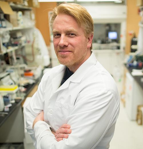
On June 17, Ajit Singh, PhD (above), Partner at Artiman Ventures, will lead a special webinar and roundtable discussion for all surgical pathologists and their practice administrators on the coming arrival of artificial intelligence-powered algorithms to aid in the primary diagnosis of certain cancers. Regulatory approval for such solutions may happen by the end of this year. Such a development would accelerate the transition from light microscopes to a fully digital pathology workflow. Singh is shown above addressing the 2018 Executive War College. (Photo copyright: The Dark Report.)
“It is now possible to do a secondary read, and even a first read, in prostate cancer with an AI system alone. In cases where there may be uncertainty, a pathologist can review the images. Now, this is specifically for prostate cancer, and I think this is a tremendous positive development for diagnostic pathways,” he added.
Use of Digital Pathology with AI-Algorithms Changes Diagnostics
Pathologists who are wedded to their light microscopes will want to pay attention to the impending arrival of a fully digital pathology system, where glass slides are converted to whole-slide images and then digitized. From that point, the surgical pathologist becomes the coach and quarterback of an individual patient’s case. The pathologist guides the AI-powered image analysis algorithms. Based on the results, the pathologist then orders supplementary tests appropriate to developing a robust diagnosis and guiding therapeutic decisions for that patient’s cancer.
In his interview with The Dark Report, Singh explained that the first effective AI-powered algorithms in digital pathology will be developed for prostate cancer and skin cancer. Both types of cancer are much less complex than, say, breast cancer. Moreover, the AI developers have decades of prostate cancer and melanoma cases where the biopsies, diagnoses, and downstream patient outcomes create a rich data base from which the algorithms can be trained and tuned.
This webinar is organized as a roundtable discussion so participants can interact with the expert panelists. The Chair and Moderator is Ajit Singh, PhD, Adjunct Professor at the Stanford School of Medicine and Partner at Artiman Ventures.
The panelists (above) represent academic pathology, community hospital pathology, and the commercial sector. They are:
Because the arrival of automated analysis of digital pathology images will transform the daily routine of every surgical pathologist, it would be beneficial for all pathology groups to have one or more of their pathologists register and participate in this critical webinar.
The roundtable discussion will help them understand how quickly AI-powered image analysis is expected be cleared for use by the FDA in such diseases as prostate cancer and melanomas. Both types of cancers generate high volumes of case referrals to the nation’s pathologists, so potential for disruption to long-standing client relationships, and the possible loss of revenue for pathology groups that delay their adoption of digital pathology, can be significant.
On the flip side, community pathology groups that jump on the digital pathology bandwagon early and with the right preparation will be positioned to build stronger client relationships, increase subspecialty case referrals, and generate additional streams of revenue that boost partner compensation within their group.
Also, because so many pathologists are working remotely, Dark Daily has arranged special group rates for pathology practices that would like their surgical pathologists to participate in this important webinar and roundtable discussion on AI-powered primary diagnosis of pathology images. Inquire at info@darkreport.com or call 512-264-7103.
Clinical-Grade Artificial Intelligence (AI) for Your Pathology Lab: What’s Ready Now, What’s Coming Soon, and How Pathologists Can Profit from Its Use PREMIUM 90-MINUTE WEBINAR WITH Q&A Held Thursday, June 17, 2021 | 1-2:30 PM EDT | $195 stream on-demand or $59 DVD BUY NOW Pathologists have learned a great deal from the “early AI digital fellow,” but have they gained a comfort level with putting AI to work in their clinical practice? Artificial intelligence is poised to transform...