Many companies want to adapt consumer wearables to monitor health conditions, including biomarkers tested by medical laboratories
Clinical laboratory managers know that wearable devices for monitoring biophysical functions or measuring biomarkers are becoming more complex and capable thanks to advances in miniaturization, informatics, software, and artificial intelligence machine learning that enable new functions to be developed and proved to be accurate.
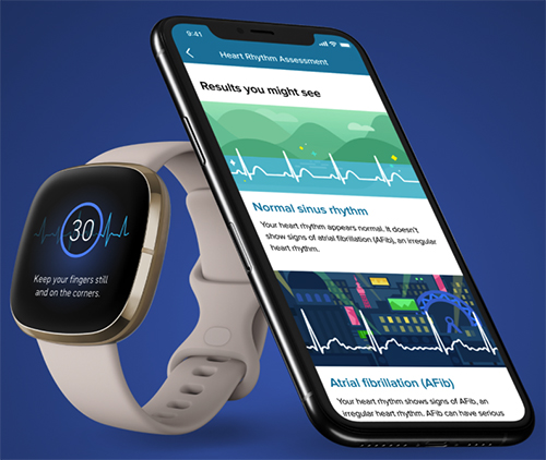
In September, Fitbit (NYSE:FIT), took that a step further. The San Francisco-based maker of personal fitness technology, “received 510(k) clearance from the US Food and Drug Administration (FDA), as well as Conformité Européenne (CE marking) in the European Union, for its electrocardiogram (ECG) app to assess heart rhythm for atrial fibrillation (AFib),” according to a press release.
The fact that Google is currently in the process of acquiring Fitbit for $2.1 billion may indicate that wearable devices to help physicians and patients diagnose and monitor health conditions will be big business in the future.
The new ECG app is available on Fitbit Sense (above), an “advanced health smartwatch.” To use the app, wearers place their finger and thumb to the stainless-steel corners on the watch and remain still for 30 seconds. The app analyzes the heart’s rhythm for signs of AFib. Individuals can take readings of their heart rhythm at any time, monitor for irregularities, and save and share the data. (Photo copyright: Fitbit.)
Helping Doctors ‘Stay Better Connected’ to Their Patients
“Helping people understand and manage their heart health has always been a priority for Fitbit, and our new ECG app is designed for those users who want to assess themselves in the moment and review the reading later with their doctor,” said Eric Friedman, Fitbit co-founder and Chief Technology Officer, in the press release.
Prior to submitting the device for approval to regulatory agencies, Fitbit conducted the clinical trial in regions throughout the US to evaluate the device’s ability to accurately detect AFib from normal sinus rhythm and generate ECG traces. The researchers proved that their algorithm was able to detect 98.7% of AFib cases (sensitivity) and was able to accurately identify normal sinus rhythms (specificity) in 100% of the cases.
Venkatesh Raman, MD, interventional cardiologist and Medical Director of the Cardiac Catheterization Lab at 609-bed MedStar Georgetown University Hospital, was Principal Investigator for the clinical study on Fitbit’s ECG app. “Physicians are often flying blind as to the day-to-day lives of our patients in between office visits. I’ve long believed in the potential for wearable devices to help us stay better connected, and use real-world, individual data to deliver more informed, personalized care,” he said in the press release.
“Given the toll that AFib continues to take on individuals and families around the world,” Raman continued, “I’m very enthusiastic about the potential of this tool to help people detect possible AFib—a clinically important rhythm abnormality—even after they leave the physician’s office.”
Fitbit ECG App Receives European CE Marking
In addition to receiving approval for the Fitbit ECG app in the US, the device also received CE marking (Conformité Européenne) for use in some European countries.
In October 2020, the app was made available to Fitbit Sense users in the US, Austria, Belgium, Czech Republic, France, Germany, Ireland, Italy, Luxembourg, the Netherlands, Poland, Portugal, Romania, Spain, Sweden, Switzerland, and the United Kingdom. The device also received approval for use in Hong Kong and India.
It is estimated that more than 33.5 million people globally have AFib, an irregular heart rhythm (arrhythmia) that can lead to stroke, blood clots, or heart failure. The American Heart Association estimates that at least 2.7 million Americans currently live with the condition. The most common symptoms experienced by those with the condition are:
Irregular heartbeat,
Heart palpitations (rapid, fluttering, quivering or pounding),
Lightheadedness,
Extreme fatigue,
Shortness of breath, and
Chest pain.
Risk factors for AFib include advancing age, high blood pressure, obesity, diabetes, European ancestry, hyperthyroidism, chronic kidney disease, alcohol use, smoking, and known heart issues such as heart failure, ischemic heart disease, and enlargement of the chambers on the left side of the heart.
According to the Centers for Disease Control and Prevention (CDC), there are more than 454,000 hospitalizations annually in the US that list AFib as the primary diagnosis. In 2018, AFib was mentioned on 175,326 death certificates with the condition being the underlying cause of death in 25,845 of those cases.
The CDC reports that cases are increasing and projects that by 2030 12.1 million people in the US will have AFib. Many people are asymptomatic of the illness and do not know they have it, which can make AFib more difficult to diagnose.
“Early detection of AFib is critical, and I’m incredibly excited that we are making these innovations accessible to people around the world to help them improve their heart health, prevent more serious conditions, and potentially save lives,” Friedman said, in a statement.
Clinical laboratory managers should monitor these developments closely. Fitbit’s FDA clearance and CE Marking of its ECG app suggest this trend is accelerating.
COMPANY OVERVIEW Digital Pathology Today is your podcast all about the world of digital pathology. Each week, we talk with industry leaders, key academics, top pathologists and more to discover the past, present and future of digital pathology. KEY AREAS OF SERVICE Provide news, information and analysis about the state of digital pathology Examine digital pathology’s past, present and future Engage key leaders in digital pathology to discuss topics of interest to our listeners KEY...
COMPANY OVERVIEW Gestalt Diagnostics provides digital pathology solutions, technical and integration services and support to pathology laboratories. Gestalt has developed PathFlow™, an enterprise software platform specifically designed to bring the benefits of digital workflow to pathologists and pathology laboratories. Gestalt has built upon its team’s experience in developing and deploying a robust radiology PACS and workflow solution that was deployed within Inland Imaging, Laboratory...
Because of ‘shelter in place’ orders, many anatomic pathologists are reviewing digital images from home during the COVID-19 outbreak and demonstrating the value of whole slide imaging, digital pathology, and CMS’ recent amended remote sign-out policy
COVID-19 is already triggering many permanent changes in the way healthcare is organized and delivered in the United States. However, not until the SARS-CoV-2 pandemic eases will the full extent of these changes become visible. This will be particularly true for anatomic pathology and the profession’s expanded use of telepathology, digital pathology, and whole-slide imaging.
Since early March, specimen referrals and revenues have collapsed at anatomic pathology groups and laboratories across the nation. Dark Daily’s sister publication, The Dark Report (TDR), was first to quantify the magnitude of this collapse in tissue referrals to pathology groups. In an interview with The Dark Report, Kyle Fetter, Executive Vice President and General Manager of Diagnostic Services at XIFIN, Inc., explained that pathology clients using XIFIN’s revenue cycle management services were seeing an average 40% decrease in specimens. And, for certain pathology sub-specialties, the drop-off in specimen referrals was as much as 90%. (See TDR, “From Mid-March, Labs Saw Big Drop in Revenue,” April 20, 2020.)
The College of American Pathologists (CAP) appealed to the Centers for Medicare and Medicaid Services (CMS) to allow pathologists to work remotely. In response, CMS issued a memorandum which stated, “Due to the public health emergency posed by COVID-19 and the urgent need to expand laboratory capacity, CMS is exercising its enforcement discretion to adopt a temporary policy of relaxed enforcement in connection with laboratories located at temporary testing sites under the conditions outlined herein.”
Since then, many physicians, including pathologists, have quickly adapted to working remotely in some form.
Push for Remote Pathology Services Acknowledges Anatomic Pathologist Shortage
The CMS memorandum (QSO-20-21-CLIA), which the federal agency issued to laboratory surveyors on March 26, 2020, notes that CMS will exercise “enforcement discretion to ensure pathologists may review pathology slides remotely” if certain defined conditions are met.
CMS’ decision, which “is applicable only during the COVID-19 public health emergency,” is intended to increase capacity by allowing remote site review of clinical laboratory data, results, and pathology slides.
Ordinarily, CLIA regulations for cytology (a branch of study that focuses on the biological structure of cells) state that cytology slide preparations must be evaluated on the premises of a laboratory that is certified to conduct testing in the subspecialty of cytology. However, a fast-acting Congressional letter sent by 37 members of Congress to US Department of Health and Human Services (HHS) Secretary Alex Azar II, MD, states, “it is unwise and unnecessary to overburden the remaining pathologists with excess work due to staffing shortages, thereby increasing the risk of burnout, medical error, and further shortages in staffing due to exposure. The number of COVID-19 cases will increase and peak over the next two months and will stretch existing healthcare systems to their limits.”
Decreasing Number of ‘Active Pathologists’ Drives Adoption of Telepathology, Digital Pathology, and Whole-slide Imaging
The current COVID-19 outbreak is just the latest factor in support of enabling remote review of anatomic pathology images and cases. The trend of using telepathology, whole-slide imaging (WSI), and digital pathology systems has been gathering momentum for several years. Powerful economic forces support this trend.
The Dark Report devoted its June 10, 2019, issue to a deep dive of the challenges currently facing the anatomic pathology profession. In particular, TDR noted a study published May 31, 2019, in the Journal of the American Medical Association (JAMA) Network Open, titled, “Trends in the US and Canadian Pathologist Workforces from 2007 to 2017.” The study’s authors—pathologists in the United States and Canada—reported that between 2007 and 2017 the number of active pathologists in the United States decreased from 15,568 to 12,839—a 17.53% decline.
TDR noted that these findings imply there are fewer pathologists in the United States today in active practice to handle the steady increase in the number of cases requiring diagnostic review. In turn, this situation could lead to delays in diagnoses detrimental to patient care.
In the United States, the COVID-19 pandemic created an “immediate need for remote sign-outs, reviews, and consults,” said Mike Bonham, MD, PhD (above), Chief Medical Officer for Proscia, a digital pathology software developer, in an interview with Dark Daily. “In the context of highly relevant workflow and workforce challenges, it reinforces the opportunity for wider adoption of digital pathology.” Prior to the outbreak of COVID-19, several distinct forces were driving adoption and use of digital pathology in combination with traditional microscopy, he said. (Photo copyright: Proscia.)
Distinct Forces Beginning to Reshape Anatomic Pathology
In recent years, the anatomic pathology profession has faced growing financial pressure, a shrinking workforce, and a surge in the global demand for pathology—issues that come at a time when biopsies and cancer diagnostics require greater expertise.
The UCSF School of Medicine started with frozen slide sections and moved to the broader volume of pathology slides. Since 2015, UCSF’s School of Medicine has moved toward a fully digital pathology operation and has serialized the adoption by specialty, according to Zoltan Laszik, MD, PhD, attending physician at UCSF and Professor of Clinical Pathology in UCSF’s Departments of Pathology and Laboratory Medicine.
Laszik is among a handful of specialists and digital pathology early adopters who collaborated on the new Dark Daily white paper, which is available for free download.
Through the adoption of digital pathology, glass slides are digitized using a whole-slide image scanner, then analyzed through image viewing software. Although the basic viewing functionality is not drastically different than that provided by a microscope, digitization does bring improvements in lab efficiency, diagnostic accuracy, image management, workflows, and revenue enhancements.
Additionally, artificial intelligence (AI)-based computational applications have emerged as an integral part of the digital pathology workflow in some settings, the white paper explains.
“These developments are important to anatomic pathologists because the traditional pathology business model continues to transform at a steady pace,” noted Robert L. Michel, Editor-in-Chief of The Dark Report.
Anthony Magliocco, MD, FRCPC, FCAP, President and CEO of Protean BioDiagnostics and former Professor and Chair of Pathology at Moffitt Cancer Center, is featured in the white paper as well. His new pathology service model provides routine pathology services, precision oncology, second opinions, liquid biopsies, genetics, and genomics to cancer centers from a Florida-based specialty laboratory.
To register for this important learning opportunity, click here or place this URL in your web browser: https://www.darkdaily.com/webinar/streamlined-operations-increased-revenue-higher-quality-of-care-conclusive-evidence-on-the-value-of-adopting-digital-pathology-in-your-lab/.
These digital pathology technologies represent an innovative movement shaping the present and future of pathology services. Pathologists wanting to learn more are encouraged to sign up for the May 13 webinar, which will build on the body of evidence and commentary that is included in the new white paper, and which will be available for free on-demand download following the live broadcast.
Previously considered “junk,” scientists learn that parts of DNA which don’t produce proteins are more important than first thought
It turns out that the long stretches of DNA in the human genome that are commonly called “junk DNA” or “dark DNA” may be doing important work. Researchers at the Ontario Institute for Cancer Research (OICR) recently published their findings about stretches of junk DNA that may have a role in how cancers develop.
Until very recently only about 2% of the human genome was considered important. Researchers were most interested in the portion of DNA that produces proteins, known as the coding region or CDS (coding sequence). The rest of the genome, 98% of it, was considered “junk” DNA. The OICR researchers found that all that DNA might not be junk after all, but instead plays a critical role in preventing cancer.
The OICR study included samples from more than 1,800 patients
with different types of cancer. The researchers looked at more than 100,000
sections of each patient’s genome and examined mutation patterns within the
large, non-coding parts of DNA. The researchers found that those non-coding
areas can control how and when certain genes are activated.
“One of the 30 key regions discovered was predicted to have a significant role in regulating a known anti-tumor gene in cancer cells, despite being more than 250,000 base pairs away from the gene in the genome,” states the news release.
Viewing DNA in New Ways Brings Insights
Within just the last few years, researchers have begun to
consider the architecture of DNA, and have begun to study it as a three-dimensional
(3D) structure. What they’ve learned is that the twisting, turning way that DNA
is packaged tightly into the nucleus of cells serves an important purpose. The
structure of DNA allows areas of non-coding DNA to be in close proximity to
other sections, as the OICR researchers discovered with the anti-tumor gene.
This discovery has revealed patterns that weren’t obvious
when the DNA was examined as if it were stretched out in a flat line. Before
scientists considered DNA in three dimensions, they were only able to identify certain
mutations, such as BRCA,
which are rare but indicate a higher cancer risk.
In looking at DNA as a whole, including the non-coding parts, researchers were able to identify specific Single Nucleotide Polymorphisms (SNPs) that when in particular positions can impact a person’s risk of cancer.
“Cancer-driver mutations are relatively rare in these large non-coding regions that often lie far from genes, presenting major challenges for systematic data analysis,” noted Jüri Reimand, PhD (above), molecular geneticist and principal investigator at OICR, Assistant Professor at the University of Toronto, and lead author of the OICR study. “Powered by novel statistical tools and whole genome sequencing data from more than 1,800 patients, we found evidence of new molecular mechanisms that may cause cancer and give rise to more-aggressive tumors.” (Photo copyright: University of Toronto.)
Another study conducted by scientist in England at Cancer Research UK and published in the British Journal of Cancer (BJC), titled, “Nongenic Cancer-Risk SNPs Affect Oncogenes, Tumour-Suppressor Genes, and Immune Function,” reached similar conclusions. The authors of that study wrote that “cancer-risk SNPs are associated with the expression levels of oncogenes [a gene with the potential to cause cancer] and tumor suppressor genes at a far greater rate than expected by chance. This indicates not only that mutations in these cancer genes are important, but also that genetic control of these genes by regulatory variants plays an important role.”
CRISPR and AI Bring New Discoveries
All of these genetic discoveries are a long way from being useful in developing diagnostics and treatments that can be used to help patients. However, researchers are using existing gene sequencing technologies such as CRISPR, along with artificial intelligence (AI), to speed up development.
The OICR researchers used CRISPR-Cas9 genome editing to explore the cancer-driving area of DNA they identified. And the researchers who conducted the BJC study plan to develop AI models based on their work that will better predict cancer risk.
“What we found surprised us, as it had never been reported before. Our results show that small genetic variations work collectively to subtly shift the activity of genes that drive cancer. We hope that this approach could one day save lives by helping to identify people at risk of cancer as well as other complex diseases,” said John Quackenbush, PhD, Professor, Computational Biology and Chair, Department of Biostatistics, Harvard T.H. Chan School of Public Health and lead author of the Cancer Research UK study, in a news release.
Clinical pathology may be on the cusp of change, driven in
large part by the discoveries being made in the realms of omics. New cancer
biomarkers coming out of these studies would be a boon to anatomic pathologists
and clinical laboratory diagnostics. Increased development of precision
medicine treatments based on these research findings could save many lives.