Ten year collaboration between Google and Harvard may lead to a deeper understanding of the brain and new clinical laboratory diagnostics
With all our anatomic pathology and clinical laboratory science, we still do not know that much about the structure of the brain. But now, scientists at Harvard University and Google Research studying the emerging field of connectomics have published a highly detailed 3D reconstruction of human brain tissue that allows visualization of neurons and their connections at unprecedented nanoscale resolutions.
Further investigation of the nano-connections within the human brain could lead to novel insights about the role specific proteins and molecules play in the function of the brain. Though it will likely be years down the road, data derived from this study could be used to develop new clinical laboratory diagnostic tests.
The data to generate the model came from Google’s use of artificial intelligence (AI) algorithms to color-code Harvard’s electron microscope imaging of a cubic millimeter of neural tissue—equivalent to a half-grain of rice—that was surgically removed from an epilepsy patient.
“That tiny square contains 57,000 cells, 230 millimeters of blood vessels, and 150 million synapses, all amounting to 1,400 terabytes of data,” according to the Harvard Gazette, which described the project as “the largest-ever dataset of human neural connections.”
“A terabyte is, for most people, gigantic, yet a fragment of a human brain—just a minuscule, teeny-weeny little bit of human brain—is still thousands of terabytes,” said neuroscientist Jeff W. Lichtman, MD, PhD, Jeremy R. Knowles Professor of Molecular and Cellular Biology, whose Lichtman Lab at Harvard University collaborated on the project with researchers from Google. The two labs have been working together for nearly 10 years on this project, the Harvard Gazette reported.
Lichtman’s lab focuses on the emerging field of connectomics, defined “as understanding how individual neurons are connected to one another to form functional networks,” said neurobiologist Wei-Chung Allen Lee, PhD, Assistant Professor of Neurology, Harvard Medical School, in an interview with Harvard Medical News. “The goal is to create connectomes—or detailed structural maps of connectivity—where we can see every neuron and every connection.” Lee was not involved with the Harvard/Google Research study.
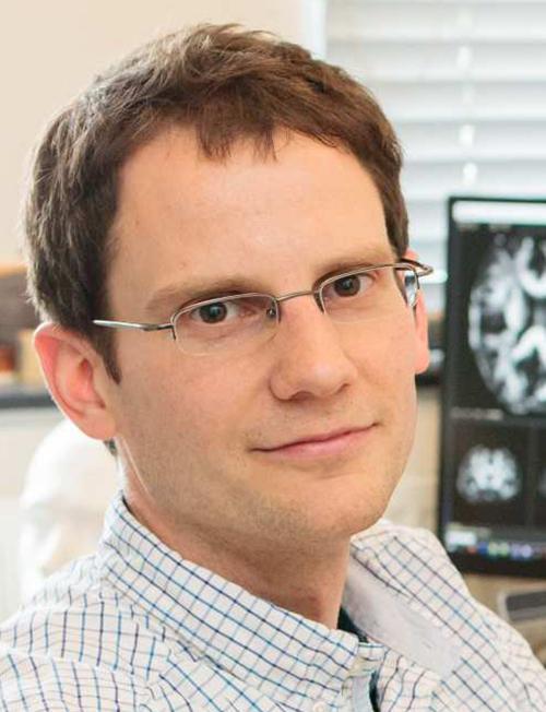
“The human brain uses no more power than a dim incandescent light bulb, yet it can accomplish feats still not possible with the largest artificial computing systems,” wrote Google Research scientist Viren Jain, PhD (above), in a blog post. “To understand how requires a level of understanding more profound than knowing what part of the brain is responsible for what function. The field of connectomics aims to achieve this by precisely mapping how each cell is connected to others.” Google’s 10-year collaboration with Harvard University may lead to new clinical laboratory diagnostics. (Photo copyright: Google Research.)
Study Data and Tools Freely Available
Along with the Science paper, the researchers publicly released the data along with analytic and visualization tools. The study noted that the dataset “is large and incompletely scrutinized,” so the scientists are inviting other researchers to assist in improving the model.
“The ability for other researchers to proofread and refine this human brain connectome is one of many ways that we see the release of this paper and the associated tools as not only the culmination of 10 years of work, but the beginning of something new,” wrote Google Research scientist Viren Jain, PhD, in a blog post that included links to the online resources.
One of those tools—Neuroglancer—allows any user with a web browser to view 3D models of neurons, axons, synapses, dendrites, blood vessels, and other objects. Users can rotate the models in xyz dimensions.
Users with the requisite knowledge and skills can proofread and correct the models by signing up for a CAVE (Connectome Annotation Versioning Engine) account.
Researchers Found Several Surprises
To perform their study, Lichtman’s team cut the neural tissue into 5,000 slices, each approximately 30 nanometers thick, Jain explained in the blog post. They then used a multibeam scanning electron microscope to capture high-resolution images, a process that took 326 days.
Jain’s team at Google used AI tools to build the model. They “stitched and aligned the image data, reconstructed the three dimensional structure of each cell, including its axons and dendrites, identified synaptic connections, and classified cell types,” he explained.
Jain pointed to “several surprises” that the reconstruction revealed. For example, he noted that “96.5% of contacts between axons and their target cells have just one synapse.” However, he added, “we found a class of rare but extremely powerful synaptic connections in which a pair of neurons may be connected by more than 50 individual synapses.”
In their Science paper, the researchers suggest that “these powerful connections are not the result of chance, but rather that these pairs had a reason to be more strongly connected than is typical,” Jain wrote in the blog post. “Further study of these connections could reveal their functional role in the brain.”
Mysterious Structures
Another anomaly was the presence of “axon whorls,” as Jain described them, “beautiful but mysterious structures in which an axon wraps itself into complicated knots.”
Because the sample came from an epilepsy patient, Jain noted that the whorls could be connected to the disease or therapies or could be found in all brains.
“Given the scale and complexity of the dataset, we expect that there are many other novel structures and characteristics yet to be discovered,” he wrote. “These findings are the tip of the iceberg of what we expect connectomics will tell us about human brains.”
The researchers have a larger goal to create a comprehensive high-resolution map of a mouse’s brain, Harvard Medical News noted. This would contain approximately 1,000 times the data found in the 1-cubic-millimeter human sample.
Dark Daily has been tracking the different fields of “omics” for years, as research teams announce new findings and coin new areas of science and medicine to which “omics” is appended. Connectomics fits that description.
Though the Harvard/Google research is not likely to lead to diagnostic assays or clinical laboratory tests any time soon, it is an example of how advances in technologies are enabling researchers to investigate smaller and smaller elements within the human body.
This AI platform has the potential to also reduce workload of radiologists, but also of anatomic pathologists and oncologists allowing them to be more productive
When the UK’s National Health Service (NHS) recently tested an artificial intelligence (AI) platform’s ability to analyze mammograms, the AI found early signs of breast cancer that “human doctors” had previously missed, the BBC reported. This level of ability by AI might soon be adapted to aid overworked anatomic pathologists and cancer doctors in the United Kingdom.
Out of 10,000 mammograms MIA analyzed, the AI platform found “tiny signs of breast cancer in 11 women” which had not been spotted during earlier examinations, the BBC noted, adding that the cancers “were practically invisible to the human eye.”
This is a significant development in AI’s role in healthcare. Anatomic pathologists and clinical laboratory leaders will note that ongoing advancements in AI are enabling technology developers to apply their solutions to assessing radiology images, as well as in whole slide imaging used in digital pathology. In the UK, use of AI, the BBC noted, may also help ease doctor’s workloads.
“This is just the beginning of our work with Kheiron,” said Ben Glocker, PhD (above), Professor in Machine Learning for Imaging at Imperial College London and Head of ML Research at Kheiron Medical, in a news release. “We are actively working on new methodologies for the safe deployment and continuous monitoring of MIA to support a US and UK rollout. We are working hard to make sure that as many women as possible will benefit from the use of this new technology within the next year.” AI tools such as MIA may soon take much of the load from anatomic pathologists and radiologists. (Photo copyright: Imperial College London.)
MIA Cloud-based AI Platform
Kheiron was founded in 2016 and MIA was named one of the seven biggest medical breakthroughs in 2023 by ABC News. A study conducted by Imperial College London in 2023 found that MIA “could significantly increase the early detection of breast cancers in a European healthcare setting by up to 13%,” according to an Imperial news release.
“The study was conducted over three phases (two pilot phases and a live roll-out). Overall across the three phases, the AI reader found 24 more cancers than the standard human reading—a 7% relative increase—and resulted in 70 more women recalled (0.28% relative increase),” the news release reported. “Of the additional recalls, six (initial pilot), 13 (extended pilot), and 11 (live use) additional cancers were found, increasing relative cancer detection rate by 13%, 10%, and 5% respectively. [The researchers] found that 83% of the additional cancers detected using MIA in real clinical practice were invasive, showing that MIA can detect cancers where early detection is particularly vital.”
Supported by Microsoft’s Azure Cloud, MIA came together over six years based on training encompassing millions of mammograms worldwide, Healthcare Digital reported.
“AI tools are generally pretty good at spotting symptoms of a specific disease if they are trained on enough data to enable them to be identified. This means feeding the program with as many different anonymized images of those symptoms as possible, from as diverse a range of people as possible,” Sarah Kerruish, Chief Strategy Officer, Kheiron, told Healthcare Digital.
MIA has been trained to “recognize subtle patterns and anomalies” that can point to “cancerous cells even in their earliest stages of development,” Dataconomy reported.
MIA Finds Early Cancer Signs
In the pilot study, MIA examined mammograms from 10,889 women. Each image had previously been reviewed by two radiologists, the BBC reported.
Findings include the following according to Healthcare Digital:
MIA “flagged” all people the physicians previously identified with symptoms.
The AI platform discovered 11 people with cancer the doctors did not identify.
The cancer MIA discovered—and the doctors did not—suggested cancer in early stages.
So, how did the doctors miss the cancer that MIA spotted? Gerald Lip, MD, Clinical Director for Breast Screening in North East Scotland who led the pilot study for the NHS, told Healthcare Digital, “part of the power of AI is it’s not prone to exhaustion or distraction.
“There is an element of fatigue,” he said. “You get disruptions, someone’s coming in, someone’s chatting in the background. There are lots of things that can probably throw you off your regular routine as well. And in those days when you have been distracted, you go, ‘how on earth did I miss that?’ It does happen.”
Lip is also the Chief Investigator in the Mammography Artificial Intelligence Project in the Industrial Center for Artificial Intelligence and Digital Diagnostics in Scotland.
“I see MIA as a friend and an augmentation to my practice,” he told Healthcare Digital. “MIA isn’t perfect. It had no access to patient history so [it] would flag cysts that had already been identified by previous scans and designated harmless.”
AI as a Safety Net
In the 2023 study, researchers from Imperial College London deployed MIA as an extra reader for mammograms of 25,065 women who visited screening sites in Hungary between April 2021 and January 2023, according to a news release.
“Our prospective real-world usage data in Hungary provides evidence for a significant, measurable increase of early breast cancer detection when MIA is used in clinical practice,” said Peter Kecskemethy, PhD, CEO and co-founder of Kheiron Medical, in the news release.
“Our study shows that AI can act as an effective safety net—a tool to prevent subtler signs of cancer from falling through the cracks,” said Ben Glocker, PhD, Professor in Machine Learning for Imaging at Imperial College London and Head of ML Research at Kheiron Medical, in the news release.
More studies are needed before MIA can be used in clinical settings. Nevertheless, use of AI in radiology—specifically mammograms—where the AI tool can identify very small cancers typically undetectable by radiologists, would be a boon to cancer doctors and the patients they treat.
So far, the research suggests that the AI-powered MIA has benefits to deployment in breast cancer screening. Eventually, it may also make impressive contributions to medical diagnosis and patient care, particularly if MIA eventually proves to be effective at analyzing the whole slide images used by anatomic pathologists.
As the cancer registry expands it will increasing become more useful to anatomic pathologists, histopathologists, oncologists, and even clinical laboratories
Oncologists, histopathologists, anatomic pathologists, and other cancer physicians now have a powerful new Wikipedia-style tumor registry to help them with their diagnoses and in educating patients on their specific types of cancer. Clinical laboratory managers may find it useful to understand the value this searchable database, and it can help their staff pathologists as well.
Free to use by both physicians and patients the World Tumor Registry (WTR) is designed “to minimize diagnostic errors by giving doctors a searchable online database of cancers that have been collected and categorized with cellular images collected from around the world,” Pittsburg-Post Gazette reported.
Prompt, accurate cancer diagnoses offer cancer patients the best chance for optimal treatment outcomes. However, many medical professionals around the globe do not have the training and resources to offer superior cancer diagnoses. That deficiency can translate to inferior treatment options and lower survival rates among cancer patients.
To help improve cancer diagnoses, pathologist Yuri E. Nikiforov, MD, PhD, Division Director, Molecular and Genomic Pathology, Vice Chair of the Department of Pathology, and Professor of Pathology, University of Pittsburgh, developed the WTR to provide educational and practical resources for individuals and organizations involved in cancer research.
Officially announced at the United States and Canadian Academy of Pathology (USCAP) annual convention, the WTR is an open-access catalog of digital microscopic images of human cancer types and subtypes.
The lower cost of technology and improved speed of access via the internet are technologies enabling this effort.
“We are creating sort of a Wikipedia for cancer images,” said Alyaksandr V. Nikitski, MD, PhD (above), Research Assistant Professor of Pathology, Division of Molecular and Genomic Pathology at Pittsburg School of Medicine and Administrative Director of the WTR, in an exclusive interview with Dark Daily. “Anyone in the world, if they can access the internet, can look at the well-annotated, diagnostic digital slides of cancer,” said Nikitski. Clinical laboratories may also find this new pathology tool useful. (Photo copyright: Alyaksandr V. Nikitski)
Minimizing Diagnostic Errors
Based in Pittsburgh, the WTR is freely available to anyone for viewing digital pathology slides of known cancer tumors as well as borderline and questionable cases. On the website, individuals can search for pictures of tumors in the registry by diagnosis, specific cohorts, and by microscopic features. Individuals may search further by tumor type and subtype to receive a picture of related tumors.
According to the WTR website, the mission of the nonprofit “is to minimize diagnostic errors, eliminate inequality in cancer recognition, diagnosis, and treatment in diverse populations, and improve outcomes by increasing access to the diagnostic pathology expertise and knowledge of microscopic characteristics of cancers that occur in different geographic, environmental, and socio-economic settings.”
This new comprehensive initiative will eventually encompass cancer images from all over the world.
“Let’s assume that I am a pathologist or a trainee who has little experience, or I don’t have access to collections of atypical tumors,” Nikitski explained. “I can view tumor collections online [in the WTR database] and check how typical and rare tumors look in various geographic regions and environmental settings.”
Once an image of a slide is selected, users will then receive a brief case history of the tumor in addition to such data as the age of the patient, their geographic location, sex, family history of the disease, and the size and stage of the tumor.
Increasing Probability of Correct Diagnosis
Pathologists and clinicians may also predict the probability of a particular diagnosis by searching under the microscopic feature of the database. This feature utilizes an innovative classifier known as PathDxFinder, where users may compare a slide from their lab to slides in the database by certain criteria. This includes:
After completing the questions above, the user presses the “predict diagnosis” button to receive the probability of cancer and most likely diagnosis based on the answers provided in the questionnaire.
WTR Editorial Boards
The WTR represents collections for each type of cancer site, such as lung or breast. A chairperson and editorial board are responsible for reviewing submitted slides before they are placed online. The editorial boards include 20 pathologists who are experts in diagnosing cancer categories, Nikitski explained.
Thousands of identified microscopic whole slide images (WSI) representing various types of cancer are deposited by the editors and other contributors to the project. The editorial board then carefully analyzes and compiles the data before posting the images for public viewing.
The editorial boards are located in five world regions:
Africa and the Middle East
Asia and Oceania
Central and South America
North America and Europe
Northern Asia
Any physicians or pathologists can contribute images to the database, by “simply selecting the editor of their region on the website, writing their name, and asking if they can submit tumor cases,” Nikitski stated.
“We have established a platform that allows pathologists to contact editors who are in the same geographic region,” he added.
Helping Physicians Identify Cancer Types
In a YouTube video, Nikiforov states that the WTR is an “educational nonprofit organization rooted in [the] beliefs that every cancer patient deserves accurate and timely diagnosis as the first and essential step in better treatment and outcomes.”
“We believe this can be achieved only when modern diagnostic tools and technologies are freely available to every physician and pathologist. Only when we understand how microscopic features of cancer vary in different geographic, environmental and ethnic populations, and only by integrating histopathology with clinical immunohistochemical and molecular genetic information for every cancer type,” he stated.
Since patient privacy is important, the database contains only basic data about patients, and all patient information is protected.
Launched in March, there are currently more than 400 thyroid tumor slides available to view in the online database. At the time of the announcement, the WTR platform was planned to be implemented in three phases:
Thyroid cancer (released in March of this year).
Lung cancer and breast cancer (anticipated to be completed by the third quarter of 2026).
Remaining cancers, including brain, soft tissue and bone, colorectal, head and neck, hematolymphoid, female genital, liver, pancreatic, prostate and male genital, skin, urinary system, pediatric, other endocrine cancers, and rare cancers (anticipated to be completed by the end of 2029).
“We believe that this resource will help physicians and pathologists practicing in small or big or remote medical centers to learn how cancer looks under a microscope in their own communities,” Nikiforov said in the video. “We also see WTR as a platform that connects physicians and scientists from different parts of the world who can work together to better understand and treat cancer.”
Catalogs like the World Tumor Registry might potentially create a pool of information that that could be mined by analytical and artificial intelligence (AI) platforms to ferret out new ways to improve the diagnosis of certain types of cancer and even enable earlier diagnoses.
“It is an extremely useful resource,” Nikitski said.
Anatomic pathologists will certainly find it so. And clinical laboratory managers may find the information useful as well when interacting with histopathologists and oncologists.
Groups representing academic publishers are taking steps to combat paper mills that write the papers and then sell authorship spots
Clinical laboratory professionals rely on peer-reviewed research to keep up with the latest findings in pathology, laboratory medicine, and other medical fields. They should thus be interested in new efforts to combat the presence of “research paper mills,” defined as “profit oriented, unofficial, and potentially illegal organizations that produce and sell fraudulent manuscripts that seem to resemble genuine research,” according to the Committee on Publication Ethics (COPE), a non-profit organization representing stakeholders in academic publishing.
“They may also handle the administration of submitting the article to journals for review and sell authorship to researchers once the article is accepted for publication,” the COPE website states.
In a recent example of how paper mills impact scholarly research, multinational publishing company John Wiley and Sons (Wiley) announced in The Scholarly Kitchen last year that it had retracted more than 1,700 papers published in journals from the company’s Hindawi subsidiary, which specializes in open-access academic publishing.
“In Hindawi’s case, this is a direct result of sophisticated paper mill activity,” wrote Jay Flynn, Wiley’s Executive Vice President and General Manager, Research, in a Scholarly Kitchen guest post. “The extent to which our processes and systems were breached required an end-to-end review of every step in the peer review and publishing process.”
In addition, journal indexer Clarivate removed 19 Hindawi journals from its Web of Science list in March 2023, due to problems with their editorial quality, Retraction Watch reported.
Hindawi later shut down four of the journals, which had been “heavily compromised by paper mills,” according to a blog post from the publisher.
Wiley also announced at that time that it would temporarily pause Hindawi’s special issues publishing program due to compromised articles, according to a press release.
“We urgently need a collaborative, forward-looking and thoughtful approach to journal security to stop bad actors from further abusing the industry’s systems, journals, and the communities we serve,” wrote Jay Flynn (above), Wiley EVP and General Manager, Research and Learning, in an article he penned for The Scholarly Kitchen. “We’re committed to addressing the challenge presented by paper mills and academic fraud head on, and we invite our publishing peers, and the many organizations that work alongside us, to join us in this endeavor.” Clinical laboratory leaders understand the critical need for accurate medical research papers. (Photo copyright: The Scholarly Kitchen.)
Using AI to Detect Paper Mill Submissions
Wiley acquired Hindawi in 2021 in a deal valued at $298 million, according to a press release, but the subsidiary has since become a financial drain for the company.
The journals earn their revenue by charging fees to authors. But in fiscal year 2024, which began last fall, “Wiley expects $35-40 million in lost revenue from Hindawi as it works to turn around journals with issues and retract articles,” Retraction Watch reported, citing an earnings call.
Wiley also revealed that it would stop using the Hindawi brand name and bring the subsidiary’s remaining journals under its own umbrella by the middle of 2024.
The service will incorporate tools to detect signs that submissions originated from paper mills, including similarities with “known papermill hallmarks” and use of “tortured phrases” indicating that passages were translated by AI-based language models, according to a press release.
These tools include:
Papermill Similarity Detection: Checks for known papermill hallmarks and compares content against existing papermills papers.
Problematic Phrase Recognition: Flags unusual alternatives to established terms.
Unusual Publication Behavior Detection: Identifies irregular publishing patterns by paper authors.
Researcher Identity Verification: Helps detect potential bad actors.
Gen-AI Generated Content Detection: Identifies potential misuse of generative AI.
Journal Scope Checker: Analyzes the article’s relevance to the journal.
The company said that the new service will be available through Research Exchange, Wiley’s manuscript submission platform, as early as next year.
Other Efforts to Spot Paper Mill Submissions
Previously, STM announced the launch of the STM Integrity Hub, with a mission “to equip the scholarly communication community with data, intelligence, and technology to protect research integrity,” Program Director Joris van Rossum, PhD, told The Scholarly Kitchen.
In 2023, the group announced that the hub would integrate Papermill Alarm from Clear Skies, a paper mill detection tool launched in 2022 with a focus on cancer research. It uses a “traffic-light rating system for research papers,” according to a press release.
In an announcement about the launch of Wiley’s Papermill Detection service, Retraction Watch suggested that one key to addressing the problem would be to reduce incentives for authors to use paper mills. Those incentives boil down to the pressure placed on many scientists, clinicians, and students to publish manuscripts, according to the research report from STM and COPE.
In one common scenario, the report noted, a paper mill will submit a staff-written paper to multiple journals. If the paper is accepted, the company will list it on a website and offer authorship spaces for sale.
“If a published paper is challenged, the ‘author’ may sometimes back down and ask for the paper to be retracted because of data problems, or they may try to provide additional supporting information including a supporting letter from their institution which is also a fake,” the report noted.
All of this serves as a warning to pathologists and clinical laboratory professionals to carefully evaluate the sources of medical journals publishing studies that feature results on areas of healthcare and lab medicine research that are of interest.
Trifecta of forces at work that will affect the clinical laboratory and pathology industries have been described as a ‘perfect storm’ requiring lab and practice managers to be well informed
Digital pathology, artificial intelligence (AI) in healthcare, and the perfect storm of changing federal regulations, took centerstage at the 29th Executive War College on Diagnostics, Clinical Laboratory, and Pathology Management in New Orleans this week, where more than 1,000 clinical laboratory and pathology leaders convened over three days.
This was the largest number of people ever onsite for what has become the world’s largest event focused exclusively on lab management topics and solutions. Perhaps the highlight of the week was the federal Food and Drug Administration’s (FDA’s) announcement of its final rule on Laboratory Developed Tests (LDTs). Overall, the conference featured more than 120 speakers, many of them national thought leaders on the topic of clinical lab and pathology management. More than 65% of the audience onsite were executive level lab managers.
“The level of interest in the annual Executive War College is testimony to the ongoing need for dynamic, engaging, and highly relevant conference events,” said Robert Michel (above), Editor-in-Chief of Dark Daily and its sister publication The Dark Report, and founder of the Executive War College. “These in-person gatherings present great opportunities for clinical laboratory and pathology managers and leaders to network and speak with people they otherwise might not meet.” (Photo copyright: Dark Intelligence Group.)
Demonstrating Clinical Value
For those who missed the action onsite, the following is a synopsis of the highlights this week.
Lâle White, Executive Chair and CEO of XiFin, spoke about the future of clinical laboratory testing and the factors reshaping the industry. There are multiple dynamics impacting healthcare economics and outcomes—namely rising costs, decreasing reimbursements, and the move to a more consumer-focused healthcare. But it is up to labs, she said, to ensure their services are not simply viewed as a commodity.
“Laboratory diagnostics have the potential to change the economics of healthcare by really gaining efficiencies,” she noted. “And it’s up to labs to demonstrate clinical value by helping physicians manage two key diagnostic decision points—what tests to order, and what to do with the results.”
But even as labs find ways to increase the value offered to clinicians, there are other disruptive factors in play. Consumer-oriented tech companies such as Google, Apple, and Amazon are democratizing access to patient data in unforeseen ways, and Medicare Advantage plans are changing the way claims are processed and paid.
Clinical labs are fundamental components of the public health infrastructure. So, the CDC plans on focusing on delivering high-quality laboratory science, supported by reliable diagnostics and informatics for disease outbreaks and exposures, and engaging with public and private sector partners.
The history of MolDX and Z-Codes were the topics discussed by Gabriel Bien-Willner, MD, PhD, Chief Medical Officer for healthcare claims and transaction processing company Palmetto GBA. Molecular testing is highly complex, and the lack of well-defined billing codes and standardization makes it difficult to know if a given test is reasonable and necessary.
Z-Codes were established to clarify what molecular testing was performed—and why—prompting payers to require both Z-Codes and Current Procedural Terminology (CPT) codes when processing molecular test claims. Medicare’s MolDX program further streamlines the claims process by utilizing expertise in the molecular diagnostics space to help payers develop coverage policies and reimbursement for these tests.
FDA Final Rule on LDT Regulation
Timothy Stenzel, MD, PhD, CEO of Grey Haven Consulting and former director of the FDA’s Office of In Vitro Diagnostics reviewed the latest updates from the FDA’s Final Rule on LDT (laboratory developed test) regulation. Prior to the FDA releasing its final rule, some experts suggested that the new regulations could result in up to 90% of labs discontinuing their LDT programs, impacting innovation, and patient care.
However, the final rule on LDTs is very different from the original proposed rule which created controversy. The final rule actually lowers the regulatory burden to the point that some labs may not have to submit their LDTs at all. The FDA is reviewing dozens of multi-cancer detection assays, some of which have launched clinically as LDTs. The agency is likely to approve those that accurately detect cancers for which there is no formal screening program.
Stenzel explained the FDA’s plan to down-classify most in vitro diagnostic tests, changing them from Class III to Class II, and exempting more than 1,000 assays from FDA review. He also discussed the highlights of the Quality Management System Regulation (QMSR). Launched in January, the QMSR bought FDA requirements in line with ISO 13485, making compliance easier for medical device manufacturers and test developers working internationally.
Looming Perfect Storm of Regulatory Changes
To close out Day 1, Michel took to the stage again with a warning to clinical laboratories about the looming “Perfect Storm” trifecta—the final FDA ruling on LDTs, Z-Code requirements for genetic testing, and updates to CLIA ’92 that could result in patient data being considered a specimen.
Laboratory leaders must think strategically if their labs are to survive the fallout, because the financial stress felt by labs in recent years will only be exacerbated by macroeconomic trends such as:
Staff shortages,
Rising costs,
Decreasing and delayed reimbursements, and
Tightening supply chains.
Lab administrators looking for ways to remain profitable and prosperous should look beyond the transactional Clinical Lab 1.0 fee-for-service model and adopt Clinical Lab 2.0, which embraces HEDIS (Healthcare Effectiveness Data and Information Set) scores and STAR ratings to offer more value to Medicare Advantage and other payers.
Wednesday’s General Session agenda was packed with information about the rise of artificial intelligence, big data, and precision medicine in healthcare. Taking centerstage on the program’s final day was Michael Simpson, President and CEO of Clinisys. Simpson gave a global perspective on healthcare data as the new driver of innovation in diagnostics and patient care.