Molecular probes designed to spot minute amounts of pathogens in biological samples may aid clinical laboratories’ speed-to-answer
Driven to find a better way to isolate minute samples of pathogens from among high-volumes of other biological organisms, researchers at Canada’s McMaster University in Hamilton, Ontario, have unveiled a bioinformatics algorithm which they claim shortens time-to-answer and speeds diagnosis of deadly diseases.
Two disease pathogens the researchers specifically targeted in their study are responsible for sepsis and SARS-CoV-2, the coronavirus causing COVID-19. Clinical laboratories would welcome a technology which both shortens time-to-answer and improves diagnostic accuracy, particularly for pathogens such as sepsis and SARS-CoV-2.
Their design of molecular probes that target the genomic sequences of specific pathogens can enable diagnosticians and clinical laboratories to spot extremely small amounts of viral and bacterial pathogens in patients’ biological samples, as well as in the environment and wildlife.
“There are thousands of bacterial pathogens and being able to determine which one is present in a patient’s blood sample could lead to the correct treatment faster when time is very important,” Zachery Dickson, a lead author of the study, told Brighter World. Dickson is a bioinformatics PhD candidate in the Department of Biology at McMaster University. “The probe makes identification much faster, meaning we could potentially save people who might otherwise die,” he added.
Sepsis is a life-threatening response to infection that leads to organ failure, tissue damage, and death in hospitals worldwide. According to Sepsis Alliance, about 30% of people diagnosed with severe sepsis will die without quick and proper treatment. Thus, a “shortcut” to identifying sepsis in its early stages may well save many lives, the McMaster researchers noted.
And COVID-19 has killed millions. Such a tool that identifies sepsis and SARS-CoV-2 in minute biological samples would be a boon to hospital medical laboratories worldwide.
“We currently need faster, cheaper, and more succinct ways to detect pathogens in human and environmental samples that democratize the hunt, and this pipeline does exactly that,” Hendrik Poinar, PhD (above), McMaster Professor of Anthropology and a lead author of the study, told Brighter World. Poinar is Director of the McMaster University Ancient DNA Center. Hospital medical laboratories could help save many lives if sepsis and COVID-19 could be detected earlier. (Graphic copyright: McMaster University.)
Is Bioinformatics ‘Shortcut’ Faster than PCR Testing?
The researchers say their probes enable a shortcut to detection—even in an infection’s early stages—by “targeting, isolating, and identifying the DNA sequences specifically and simultaneously.”
The probes’ design makes possible simultaneous targeted capture of diverse metagenomics targets, Biocompare explained.
But is it faster than PCR (polymerase chain reaction) testing?
The McMaster scientists were motivated by the “challenges of low signal, high background, and uncertain targets that plague many metagenomic sequencing efforts,” they noted in their paper.
They pointed to challenges posed by PCR testing, a popular technique used for detection of sepsis pathogens as well as, more recently, for SARS-CoV-2, the coronavirus causing COVID-19.
“The (PCR) technique relies on primers that bind to nucleic acid sequences specific to an organism or group of organisms. Although capable of sensitive, rapid detection and quantification of a particular target, PCR is limited when multiple loci are targeted by primers,” the researchers wrote in Cell Reports Methods.
According to LabMedica, “A wide array of metagenomic study efforts are hampered by the same challenge: low concentrations of targets of interest combined with overwhelming amounts of background signal. Although PCR or naive DNA capture can be used when there are a small number of organisms of interest, design challenges become untenable for large numbers of targets.”
Detecting Pathogens Faster, Cheaper, and More Accurately
As part of their study, researchers tested two probe sets:
one to target bacterial pathogens linked to sepsis, and
another to detect coronaviruses including SARS-CoV-2.
They were successful in using the probes to capture a variety of pathogens linked to sepsis and SARS-CoV-2.
“We validated HUBDesign by generating probe sets targeting the breadth of coronavirus diversity, as well as a suite of bacterial pathogens often underlying sepsis. In separate experiments demonstrating significant, simultaneous enrichment, we captured SARS-CoV-2 and HCoV-NL63 [Human coronavirus NL 63] in a human RNA background and seven bacterial strains in human blood. HUBDesign has broad applicability wherever there are multiple organisms of interest,” the researchers wrote in Cell Reports Methods.
The findings also have implications to the environment and wildlife, the researchers noted.
Of course, more research is needed to validate the tool’s usefulness in medical diagnostics. The McMaster University researchers intend to improve HUBDesign’s efficiency but note that probes cannot be designed for unknown targets.
Nevertheless, the advanced application of novel technologies to diagnose of sepsis, which causes 250,000 deaths in the US each year, according to the federal Centers for Disease Control and Prevention, is a positive development worth watching.
The McMaster scientists’ discoveries—confirmed by future research and clinical studies—could go a long way toward ending the dire effects of sepsis as well as COVID-19. That would be a welcome development, particularly for hospital-based laboratories.
Several young companies hope to expand the direct-to-consumer test market by introducing new diagnostic tests to serve the women’s health market
Providing women with at-home lab test kits is the goal of a growing class of start-up companies that are bringing to market consumer test kits for a range of health conditions common to women. These companies believe they can shift a substantial volume of such testing away from the nation’s medical laboratories.
Moreover, diagnostic startups that develop at-home direct-to-consumer (DTC) clinical laboratory genetic tests have been hot commodities among venture capitalists and other healthcare investors willing to put tens of millions of dollars into these new firms. The New York Times observed that, until recently, women’s healthcare needs have rarely been the focus of new diagnostic testing companies, but that the situation may be changing.
“Femtech” (short for female technology) products and services that address the health and wellness needs of women is the new buzz word in healthcare. It describes female-focused diagnostic startups aiming at vaginal health and other medical issues that go beyond reproductive health concerns.
This, however, is a dual-edged sword for clinical laboratory leaders. Growth in this segment could lead to new diagnostics tests that boost a medical lab’s bottom line or, conversely, it could reduce revenue as patients self-diagnose urinary tract infections (UTIs), yeast infections, and other conditions through at-home DTC testing.
“The market potential is huge,” Michelle Tempest, MD (above), a partner at the London-based healthcare consulting firm Candesic, told The New York Times. “There’s definitely an increasing appetite for anything in the world which is technology and a realization that female consumer power has arrived—and that it’s arrived in healthcare.” Tempest maintains the women’s health marketplace is ripe for growth, which could mean a boon for clinical laboratory testing and diagnostics designed specifically for women. (Photo copyright: Candesic.)
Vaginal Microbiome At-home Clinical Laboratory Tests in High Demand
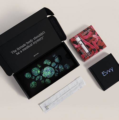
One area in particular drawing the attention of several female-led startups is vaginal health. According to an article in Vogue, test developers Juno Bio and Evvy are leading the way with at-home vaginal microbiome tests that let users “know what’s up down there.”
New York City-based Evvy ($129 for a single test or $99 each for four tests per year) uses metagenomic sequencing to identify the bacteria and fungi present in the vaginal microbiome. This information helps customers to understand their levels of protective and disruptive bacteria, which can be associated with everything from reoccurring infections and transmission of sexually transmitted diseases to infertility.
London-based Juno Bio ($149 per test) does not disclose its testing method. It does, however, provide users with a “full vaginal microbiome profile.” The profile is accessed online within a “few days” of returning the vaginal swab sample to the company’s clinical laboratory.
Both companies note that their tests are intended to be used for wellness purposes and are not meant to diagnose or treat disease or substitute for a physician’s consultation.
Gynecologist Oluwatosin Goje, MD, MSCR, FACOG, a reproductive infectious disease specialist at Cleveland Clinic, believes the availability of at-home vaginal microbiome testing will provide valuable information to both women and their doctors.
“It’s a powerful tool because it enables us to look at the entire microbial community through metagenomics and decipher how the overall composition might be affecting symptoms and infections, as well as determine the best treatment pathway,” Goje, an Evvy Medical Advisor, told Vogue. “Understanding the complete vaginal microbiome allows us to be good antibiotic stewards and only administer antibiotics when needed. Patients can also retest remotely to understand how antibiotics and other treatments impacted their vaginal microbiome.”
Evvy, which offers women an at-home vaginal microbiome test (above) that can provide insights into chronic vaginal infections and proclivity to contract sexually transmitted diseases and other women’s health issues, is one of several women-led diagnostic start-ups focused on women’s health. (Photo copyright: Evvy.)
Removing the Discomfort of Shopping for Women’s Health Products
Jamie Norwood and Cynthia Plotch, co-founders of Stix, a supplier of women’s health products and education, launched their company with a product line of at-home pregnancy and ovulation tests. They have since expanded their offerings to include urinary tract infection (UTI) and yeast infection testing and treatments.
“You can test, relieve, treat, and help prevent future infections—all from the comfort of your own home,” Norwood, told Vogue. She emphasized that this is the kind of experience healthcare consumers are demanding in today’s ever-growing direct-to-consumer clinical laboratory testing landscape. “Agonizing over confusing over-the-counter products in the drugstore aisles, or bending over backwards to pick up a prescription at the pharmacy, just isn’t cutting it for Millennial and Gen Z consumers.”
According to WebMD, yeast infections are a chronic problem for many women. While 75% of women will get at least one yeast infection in their lifetime, up to 8% get more than four a year. In addition, the federal Centers for Disease Control and Prevention (CDC) points out that bacterial vaginosis is the most common vaginal condition in females ages 15-44.
Lola Priego, is CEO and founder of blood test company Base, which sells at-home saliva and finger-prick blood tests to monitor hormone levels, vitamin levels, neurotransmitters, and blood cell markers to improve everything from sleep and diet to sex drive. She predicts direct-to-consumer testing will become as common as fitness watches.
“Eventually, at-home lab testing will be another readily-used tool, similar to your health-tracking wearables, that helps us optimize for a well-rounded healthy lifestyle in a more individualized way,” Priego told Vogue.
Femtech a ‘Significantly Underdeveloped’ Market
In its latest Analyst Note, financial data firm PitchBook maintained that the market for female health products is poised for growth. TechCrunch, which reviewed PitchBook’s analysis of female-focused health products, reported that Femtech remains a “significantly underdeveloped” slice of health-tech spending.
While women spend an estimated $500 billion annually on medical expenses, only 4% of research and development money is targeted at women’s health, PitchBook noted. In its analysis, Pitchbook predicted the global market for female-focused health products will reach $3 billion by the end of 2030. By comparison, that segment of the healthcare market totaled $820.6 million last year.
“While we still view Femtech as a niche industry, we believe secular drivers could help propel new growth opportunities in the space,” PitchBook analysts wrote. “These include the increasing representation of women in the venture-backed technology community, rising awareness and acceptance of women’s health issues, and the growing prevalence of infectious diseases among women in some countries in Africa and Asia.
“Furthermore, while the majority of Femtech products have traditionally focused on reproductive health, we believe new approaches to women’s health research will help open the door to new products and services,” they noted.
Clinical laboratory leaders will be wise to carefully watch the growth of at-home DTC tests and products targeted at female healthcare consumers since fewer trips to physicians’ offices may mean fewer test orders for local labs.
At the same time, the opportunity exists for innovative pathologists and lab managers to develop digital services that allow consumers who are self-testing to store their home-test results in the lab’s app. They can then receive relevant insights from clinical pathologists to help them fully understand the implications of the test results.
Newly combined digital pathology, artificial intelligence (AI), and omics technologies are providing anatomic pathologists and medical laboratory scientists with powerful diagnostic tools
Add “spatial transcriptomics” to the growing list of “omics” that have the potential to deliver biomarkers which can be used for earlier and more accurate diagnoses of diseases and health conditions. As with other types of omics, spatial transcriptomics might be a new tool for surgical pathologists once further studies support its use in clinical care.
Among this spectrum of omics is spatial transcriptomics, or ST for short.
Spatial Transcriptomics is a groundbreaking and powerful molecular profiling method used to measure all gene activity within a tissue sample. The technology is already leading to discoveries that are helping researchers gain valuable information about neurological diseases and breast cancer.
Marriage of Genetic Imaging and Sequencing
Spatial transcriptomics is a term used to describe a variety of methods designed to assign cell types that have been isolated and identified by messenger RNA (mRNA), to their locations in a histological section. The technology can determine subcellular localization of mRNA molecules and can quantify gene expression within anatomic pathology samples.
In “Spatial: The Next Omics Frontier,” Genetic Engineering and Biotechnology News (GEN) wrote, “Spatial transcriptomics gives a rich, spatial context to gene expression. By marrying imaging and sequencing, spatial transcriptomics can map where particular transcripts exist on the tissue, indicating where particular genes are expressed.”
In an interview with Technology Networks, George Emanuel, PhD, co-founder of life-science genomics company Vizgen, said, “Spatial transcriptomic profiling provides the genomic information of single cells as they are intricately spatially organized within their native tissue environment.
“With techniques such as single-cell sequencing, researchers can learn about cell type composition; however, these techniques isolate individual cells in droplets and do not preserve the tissue structure that is a fundamental component of every biological organism,” he added.
“Direct spatial profiling the cellular composition of the tissue allows you to better understand why certain cell types are observed there and how variations in cell state might be a consequence of the unique microenvironment within the tissue,” he continued. “In this way, spatial transcriptomics allows us to measure the complexity of biological systems along the axes that are most relevant to their function.”
“Although spatial genomics is a nascent field, we are already seeing broad interest among the community and excitement across a range of questions, all the way from plant biology to improving our understanding of the complex interactions of the tumor microenvironment,” George Emanuel, PhD (above), told Technology Networks. Oncologists, anatomic pathologists, and medical laboratory scientists my soon see diagnostics that take advantage of spatial genomics technologies. (Photo copyright: Vizgen.)
According to 10x Genomics, “spatial transcriptomics utilizes spotted arrays of specialized mRNA-capturing probes on the surface of glass slides. Each spot contains capture probes with a spatial barcode unique to that spot.
“When tissue is attached to the slide, the capture probes bind RNA from the adjacent point in the tissue. A reverse transcription reaction, while the tissue is still in place, generates a cDNA [complementary DNA] library that incorporates the spatial barcodes and preserves spatial information.
“Each spot contains approximately 200 million capture probes and all of the probes in an individual spot share a barcode that is specific to that spot.”
“The highly multiplexed transcriptomic readout reveals the complexity that arises from the very large number of genes in the genome, while high spatial resolution captures the exact locations where each transcript is being expressed,” Emanuel told Technology Networks.
Spatial Transcriptomics for Breast Cancer and Neurological Diagnostics
In that paper, the authors wrote “we envision that in the coming years we will see simplification, further standardization, and reduced pricing for the ST protocol leading to extensive ST sequencing of samples of various cancer types.”
Spatial transcriptomics is also being used to research neurological conditions and neurodegenerative diseases. ST has been proven as an effective tool to hunt for marker genes for these conditions as well as help medical professionals study drug therapies for the brain.
“You can actually map out where the target is in the brain, for example, and not only the approximate location inside the organ, but also in what type of cells,” Malte Kühnemund, PhD, Director of Research and Development at 10x Genomics, told Labiotech.eu. “You actually now know what type of cells you are targeting. That’s completely new information for them and it might help them to understand side effects and so on.”
The field of spatial transcriptomics is rapidly moving and changing as it branches out into more areas of healthcare. New discoveries within ST methodologies are making it possible to combine it with other technologies, such as Artificial Intelligence (AI), which could lead to powerful new ways oncologists and anatomic pathologists diagnose disease.
“I think it’s going to be tricky for pathologists to look at that data,” Kühnemund said. “I think this will go hand in hand with the digital pathology revolution where computers are doing the analysis and they spit out an answer. That’s a lot more precise than what any doctor could possibly do.”
Spatial transcriptomics certainly is a new and innovative way to look at tissue biology. However, the technology is still in its early stages and more research is needed to validate its development and results.
Nevertheless, this is an opportunity for companies developing artificial intelligence tools for analyzing digital pathology images to investigate how their AI technologies might be used with spatial transcriptomics to give anatomic pathologists a new and useful diagnostic tool.
The discovery is yet another factor that must be considered when developing a liquid biopsy test clinical laboratories can use to detect cancer
How often do disruptive elements present in Liquid biopsies result in misdiagnoses and unhelpful drug therapies for cancer? Researchers at the University of Washington School of Medicine (UW Medicine) in Seattle wanted to know. And the results of their study provide another useful insight for pathologists about the elements that circulate in human blood which must be understood so that liquid biopsy tests can be developed that are not affected by that factor.
Based on their case series study of 69 men with advanced prostate cancer, the UW Medicine researchers determined that 10% of men have a clonal hematopoiesis of indeterminate potential (CHIP) that can “interfere” with liquid biopsies and cause incorrect reports and unneeded prostate cancer treatment, according to their paper published in the journal JAMA Oncology.
The UW Medicine researchers advised testing for “variants in the cell-free DNA (cfDNA)” shed in blood plasma to enable appropriate treatment for people with already diagnosed prostate cancer, noted to a UW Medicine news release.
According to pathologist Colin Pritchard, MD, PhD, Associate Professor of Laboratory Medicine and Pathology at the UW Medicine, who led the research team, “clonal hematopoiesis can interfere with liquid biopsies. For example, mutations in the genes BRCA1, BRCA2, and ATM have been closely linked to cancer development.
“The good news is that, by looking at the blood cellular compartment, you can tell with pretty good certainty whether something is cancer, or something is hematopoiesis,” he said in the news release.
What Does CHIP Interference Mean to a Clinical Laboratory Blood Test?
In their published study, the UW Medicine researchers stressed the “urgent need to understand cfDNA testing performance and sources of test interferences” in light of recent US Food and Drug Administration (FDA) clearance of two PARP inhibitors (PARPi) for prostate cancer:
“We found that a strikingly high proportion of DNA repair gene variants in the plasma of patients with advanced prostate cancer are attributable to CHIP,” the researchers wrote. “The CHIP variants were strongly correlated with increased age, and even higher than expected by age group.
“The high rate of CHIP may also be influenced by prior exposure to chemotherapy,” they added. “We are concerned that CHIP interference is causing false-positive cfDNA biomarker assessments that may result in patient harm from inappropriate treatment, and delays in delivering alternative effective treatment options.
“Without performing a whole-blood control, seven of 69 patients (10%) would have been misdiagnosed and incorrectly deemed eligible for PARP-inhibitor therapy based on CHIP interference in plasma. In fact, one patient in this series had a BRCA2 CHIP clone that had been previously reported by a commercial laboratory testing company with the recommendation to use a PARPi. To mitigate these risks, cfDNA results should be compared to results from whole-blood control or tumor tissue,” the researchers concluded.
To find the clinically relevant CHIP interference in prostate cancer cfDNA testing, researchers used the UW-OncoPlex assay (developed and clinically available at UW Medicine). The assay is a multiplexed next-generation sequencing panel aimed at detecting mutations in tumor tissues in more than 350 genes, according to the UW Medicine Laboratory and Pathology website.
“To improve cfDNA assay performance, we developed an approach that simultaneously analyzes plasma and paired whole-blood control samples. Using this paired testing approach, we sought to determine to what degree CHIP interferes with the results of prostate cancer cfDNA testing,” the researchers wrote in JAMA Oncology.
Men May Receive Unhelpful Prostate Cancer Drug Therapies
The research team studied test results from 69 men with advanced prostate cancer. They analyzed patients’ plasma cfDNA and whole-blood control samples.
Tumor sequencing enabled detection of germline (cells relating to preceding cells) variants from CHIP clones.
The UW Medicine study suggested CHIP variants “accounted for almost half of the somatic (non-germline) DNA repair mutations” detected by liquid biopsy, according to the news release.
> “About half the time when the plasma is thought to contain a mutation that would guide therapy with these drugs, it actually contains CHIP variants, not prostate cancer DNA variants. That means that in about half of those tested, a patient could be told that he should be administered a drug that is not indicated to treat to his cancer,” said Colin Pritchard, MD, PhD, pathologist and Associate Professor of Laboratory Medicine and Pathology at UW Medicine in the new release. (Photo copyright: University of Washington School of Medicine.)
Other detailed findings of the UW Medicine Study:
CHIP variants of 2% or more were detected in cfDNA from 13 of 69 men.
Seven men, or 10%, having advanced prostate cancer “had CHIP variants in DNA repair genes used to determine PARPi candidacy.
CHIP variants rose with age: 0% in those 40 to 50; 12.5% in men 51 to 60; 6.3% in those 61 to 70; 20.8% in men 71 to 80; and 71% in men 81 to 90.
Whole-blood control made it possible to distinguish prostate cancer variants from CHIP interference variants.
“Men with prostate cancer are at high risk of being misdiagnosed as being eligible for PARPi therapy using current cfDNA tests; assays should use a whole-blood control sample to distinguish CHIP variants from prostate cancer,” the researchers wrote in JAMA Oncology.
Liquid Biopsies Are ‘Here to Stay’
Surgical oncologist William Cance, MD, Chief Medical and Scientific Officer, American Cancer Society (ACS) in Atlanta, recognizes the challenge of tumor biology to liquid biopsies.
“Genetic abnormalities are only one piece of the puzzle. We need to look comprehensively at tumors for the best therapy, from their metabolic changes and protein signatures in the blood to the epigenetic modifications that may occur, as cancers take hold,” he told Oncology Times. “It’s not just shed DNA in the blood.”
The UW Medicine study demonstrates the importance of understanding how all elements in liquid biopsies interact to affect clinical laboratory test results.
“I think liquid biopsies are here to stay,” Cance told Oncology Times. “They’re all part of precision medicine, tailored to the individual.”
One of the world’s fastest growing medical laboratory companies in India is using digital pathology systems and AI to replace older diagnostic technologies
Artificial intelligence (AI) is gaining acceptance around the world and use of AI to analyze digital pathology images is expected to be a major disruptor to the profession of anatomic pathology. Internationally, several pathology companies already use AI-powered solutions to diagnose cancer.
One such example is Neuberg Diagnostics, a fast-growing clinical laboratory company in Chennai, India. Neuberg has been using AI to review digital pathology images for several years, according to Chairman and Managing Director GSK Velu, PhD, BPharm.
“We already use AI in our laboratories,” Velu said in an exclusive interview with Dark Daily. “Our main reference laboratories currently use digital pathology systems to support the pathologists and many of them are using AI with these digital pathology systems.
“AI and data analytics tools are being used in other departments too, such as in our wellness department where we use AI for predictive analytics,” he added. “We also use AI in our genomics division, and we are introducing AI into other divisions slowly and steadily.”
Neuberg operates 120 laboratories in an extensive network in India, South Africa, and the United Arab Emirates (UAE), and now in the US as well.
“Our idea is to enhance the access and affordability for next-generation techniques, meaning molecular diagnostics, genomics, pathology, digital pathology, proteomics, metabolomics, and all that. This is the spirit behind Neuberg Diagnostics,” said GSK Velu, PhD, BPharm (above), Chairman and Managing Director of Neuberg Diagnostics, in an exclusive interview with The Dark Report. Clinical laboratories that are considering investing in digital pathology technologies may want to follow its development at Neuberg’s Centre for Genomic Medicine in Raleigh, NC, which opened in May. (Photo copyright: Neuberg Diagnostics.)
Replacing Older Pathology Technologies
As has been happening at other anatomic pathology centers around the world, Neuberg has been using digital pathology systems to replace older technologies. “One of our largest labs is our Bangalore Reference Lab,” Velu said. “There, we do not use microscopes for histopathology, and that lab has used digital pathology for routine review of specimens for several years now.
“But because artificial intelligence is still emerging, we can’t rely on AI with all of our digital pathology systems,” he added. “Although, of course, AI is certainly an aid to everything we do with digital pathology.
“For a variety of reasons, the adaptation of artificial intelligence in anatomic pathology is not happening as effectively nor as fast as we would like,” he noted. “So, for now, we need to wait and watch a bit longer, either because adaptation by pathologists is slow, or because AI tools are still a bit of a worry for some pathologists.
Younger Pathologists Adapt Faster to Digital Pathology
One reason could be that conventional pathologists worry about relying completely on AI for any diagnosis, Velu noted. “I’m certain that the more recent generation of pathologists who are now in their 30s, and the new people coming into pathology, will start adapting more quickly to digital pathology and to AI faster than the older generation of pathologists have done.
“The younger pathologists have a greater appreciation for the potential of digital pathology, while the older pathologists don’t want to let go of conventional diagnosis methods,” he added.
“For example, we have not yet seen where pathologists are reviewing breast image scans,” he commented. “But, at the same time, AI has been well-accepted among radiologists who are reviewing breast mammography scans.”
In India and in other markets worldwide, radiologists have adapted AI tools for breast mammography scans to diagnose breast cancer, he noted. “But that’s not happening even among pathologists who are doing cancer screening,” he said.
Velu suggested that another reason for the slow adoption of AI tools in pathology is that these systems are relatively new to the market. “Maybe the AI tools that are used with digital pathology are not as reliable as we hoped they would be, or they are not fully robust at the moment,” he speculated. “That’s why I say it will take some time before the use of AI for diagnosis becomes more widespread among pathologists. So, for now, we must wait until digital pathology and AI tools work together more seamlessly.
Replacing Conventional Pathology Technologies and Methods
“When those two technologies—AI and digital pathology systems—are linked more closely, their use will take hold in a substantial way,” Velu predicted. “When that happens, they are likely to replace conventional pathology methods completely.
“Currently, we are in the early stages of a transformation,” he added. “In our labs, you can see that the transformation is ongoing. We are using digital pathology systems even in our smaller labs. Then, the staff in our smaller labs do the processing of slides to convert them to digital images and send them to our labs in the larger cities. There, the professional staff uses AI to review those digital images and issue reports based on those images.
“Using our digital pathology systems and AI in that way means that we can make that technology available even in smaller towns and villages that have access only to our smaller labs,” he commented.
Velu added that wider use of digital pathology systems could improve the quality of care that pathologists deliver to patients in a significant way, particularly in rural areas. “Here in India, we are not seeing a huge shortage of pathologists, except in rural areas and villages,” he explained. “In those places, we could run short of pathologists.
“That is the reason we are trying to adapt the use of telepathology more widely,” he noted. “To do that, we might have technicians and histologists who will do just processing of slides so that they can send the digital images to our pathologists located in larger cities. Then, those surgical pathologists will review the cases and send the reports out. That’s the model that we are trying to slowly follow here.”
As use of digital pathology images increased, many predicted that specimens would flow from the US to India. This would happen because of the belief that the lower cost of surgical pathology in India would successfully draw business away from pathology groups here in the United States.
However, Neuberg turned the tables on that belief when it announced the opening of its Neuberg Centre for Genomic Medicine (NCGM), a state-of-the-art esoteric and genetic testing laboratory in Raleigh, NC. The NCGM lab is CLIA-certified and Neuberg says it is ready to compete with labs in this country on their home turf.
These are reasons why pathologists and pathology practice administrators in the United States may want to watch how Neuberg Diagnostics continues to develop its use of digital pathology platforms and AI-powered digital image analysis tools throughout its international network of laboratories.