Jul 20, 2018 | Digital Pathology, Instruments & Equipment, Laboratory Instruments & Laboratory Equipment, Laboratory Management and Operations, Laboratory News, Laboratory Operations, Laboratory Pathology, Laboratory Testing
Should greater attention be given to protein damage in chronic diseases such as Alzheimer’s and diabetes? One life scientist says “yes” and suggests changing how test developers view the cause of age-related and degenerative diseases
DNA and the human genome get plenty of media attention and are considered by many to be unlocking the secrets to health and long life. However, as clinical laboratory professionals know, DNA is just one component of the very complex organism that is a human being.
In fact, DNA, RNA, and proteins are all valid biomarkers for medical laboratory tests and, according to one life scientist, all three should get equal attention as to their role in curing disease and keeping people healthy.
Along with proteins and RNA, DNA is actually an “equal partner in the circle of life,” wrote David Grainger, PhD, CEO of Methuselah Health, in a Forbes opinion piece about what he calls the “cult of DNA-centricity” and its relative limitations.
Effects of Protein Damage
“Aging and age-related degenerative diseases are caused by protein damage rather than by DNA damage,” explained Grainger, a Life Scientist who studies the role proteins play in aging and disease. “DNA, like data, cannot by itself do anything. The data on your computer is powerless without apps to interpret it, screens and speakers to communicate it, keyboards and touchscreens to interact with it.”
“Similarly,” he continued, “the DNA sequence information (although it resides in a physical object—the DNA molecule—just as computer data resides on a hard disk) is powerless and ethereal until it is translated into proteins that can perform functions,” he points out.
According to Grainger, diseases such as cystic fibrosis and Duchenne Muscular Dystrophy may be associated with genetic mutation. However, other diseases take a different course and are more likely to develop due to protein damage, which he contends may strengthen in time, causing changes in cells or tissues and, eventually, age-related diseases.
“Alzheimer’s disease, diabetes, or autoimmunity often take decades to develop (even though your genome sequence has been the same since the day you were conceived); the insidious accumulation of the damaged protein may be very slow indeed,” he penned.
“But so strong is the cult of DNA-centricity that most scientists seem unwilling to challenge the fundamental assumption that the cause of late-onset diseases must lie somewhere in the genome,” Grainger concludes.
Shifting Focus from Genetics to Proteins
Besides being CEO of Methuselah Health, Grainger also is Co-Founder and Chief Scientific Advisor at Medicxi, a life sciences investment firm that backed Methuselah Health with $5 million in venture capital funding for research into disease treatments that focus on proteins in aging, reported Fierce CEO.
Methuselah Health, founded in 2015 in Cambridge, UK, with offices in the US, is reportedly using post-translational modifications for analysis of many different proteins.

“At Methuselah Health, we have shifted focus from the genetics—which tells you in an ideal world how your body would function—to the now: this is how your body functions now and this is what is going wrong with it. And that answer lies in the proteins,” stated Dr. David Grainger (above), CEO of Methuselah Health, in an interview with the UK’s New NHS Alliance. Click on this link to watch the full interview. [Photo and caption copyright: New NHS Alliance.]
This is how Methuselah Health analyzes damaged proteins using mass spectrometry, according to David Mosedale, PhD, Methuselah Health’s Chief Technology Officer, in the New NHS Alliance story:
- Protein samples from healthy individuals and people with diseases are used;
- Proteins from the samples are sliced into protein blocks and fed slowly into a mass spectrometer, which accurately weighs them;
- Scientists observe damage to individual blocks of proteins;
- Taking those blocks, proteins are reconstructed to ascertain which proteins have been damaged;
- Information is leveraged for discovery of drugs to target diseases.
Mass spectrometry is a powerful approach to protein sample identification, according to News-Medical.Net. It enables analysis of protein specificity and background contaminants. Interactions among proteins—with RNA or DNA—also are possible with mass spectrometry.
Methuselah Health’s scientists are particularly interested in the damaged proteins that have been around a while, which they call hyper-stable danger variants (HSDVs) and consider to be the foundation for development of age-related diseases, Grainger told WuXi AppTec.
“By applying the Methuselah platform, we can see the HSDVs and so understand which pathways we need to target to prevent disease,” he explained.
For clinical laboratories, pathologists, and their patients, work by Methuselah Health could accelerate the development of personalized medicine treatments for debilitating chronic diseases. Furthermore, it may compel more people to think of DNA as one of several components interacting that make up human bodies and not as the only game in diagnostics.
—Donna Marie Pocius
Related Information:
The Cult of DNA-Centricity
Methuselah Health CEO David Grainger Out to Aid Longevity
VIDEO: Methuselah Health, Addressing Diseases Associated with Aging
Understanding and Slowing the Human Aging Clock Via Protein Stability
Using Mass Spectrometry for Protein Complex Analysis
Jul 16, 2018 | Laboratory News, Laboratory Operations, Laboratory Testing, News From Dark Daily
This potential new source of diagnostic biomarkers could give clinical labs a new tool to diagnose disease earlier and with greater accuracy
Clinical laboratories may soon have a new “omics” in their toolkit and vocabulary. In addition to genomics and proteomics, anatomic pathologists could also be using “interactomics” to diagnose disease earlier and with increased accuracy.
At least that’s what researchers at ETH Zurich (ETH), an international university for technology and natural sciences, have concluded. They published the results of their study in Cell.
“Here, we present a chemoproteomic workflow for the systematic identification of metabolite-protein interactions directly in their native environments,” the researchers wrote. “Our data reveal functional and structural principles of chemical communication, shed light on the prevalence and mechanisms of enzyme promiscuity, and enable extraction of quantitative parameters of metabolite binding on a proteome-wide scale.”
Interactomics address interactions between proteins and small molecules, according to an article published in Technology Networks. The terms “interactomics” and “omics” were inspired by research that described, for the first time, the interactions and relationships of all proteins and metabolites (A.K.A, small molecules) in the whole proteome.
Medical laboratories and anatomic pathologists have long understood the interactions among proteins, or between proteins and DNA or RNA. However, metabolite interactions with packages of proteins are not as well known.
These new omics could eventually be an important source of diagnostic biomarkers. They may, one day, contribute to lower cost clinical laboratory testing for some diseases, as well.
Metabolite-Protein Interactions are Key to Cellular Processes
The ETH researchers were motivated to explore the interplay between small molecules and proteins because they have important responsibilities in the body. These cellular processes include:
“Metabolite-protein interactions control a variety of cellular processes, thereby playing a major role in maintaining cellular homeostasis. Metabolites comprise the largest fraction of molecules in cells. But our knowledge of the metabolite-protein interaction lags behind our understanding of protein-protein or protein-DNA interactomes,” the researchers wrote in Cell.
Leveraging Limited Proteolysis and Mass Spectrometry
The researchers used limited proteolysis (LiP) technology with mass spectrometry to discover metabolite-protein interactions. Results aside, experts pointed out that the LiP technology itself is significant.
“It is one of the few methods that enables the unbiased and proteome-wide profiling of protein conformational changes resulting from interaction of proteins with compounds,” stated a Biognosys blog post.
Biognosys, a proteomics company founded in 2008, was originally part of a lab at ETH Zurich.
The ETH team focused on the E. coli bacterial cell in particular and how its proteins and enzymes interact with metabolites.

“Although the metabolism of E. coli and associated molecules is already very well known, we succeeded in discovering many new interactions and the corresponding binding sites,” Paola Picotti, PhD, Professor of Molecular Systems Biology at ETH Zurich, who led the research, told Technology Networks. “The data that we produce with this technique will help to identify new regulatory mechanisms, unknown enzymes and new metabolic reactions in the cell,” she concluded. (Photo copyright: ETH Zurich.)
More than 1,000 New Interactions Discovered
The study progressed as follows, according to Technology Networks’ report:
- “Cellular fluid, containing proteins, was extracted from bacterial cells;
- “A metabolite was added to each sample;
- “The metabolite interacted with proteins;
- “Proteins were cut into smaller pieces by molecular scissors (A.K.A., CRISPR-Cas9);
- “Protein structure was altered when it interacted with a metabolite;
- “A different set of peptides emerged when the “molecular scissors” cut at different sites;
- “Pieces of samples were measured with a mass spectrometer;
- “Data were obtained, fed into a computer, and structural differences and changes were reconstructed;
- “1,650 different protein-metabolite interactions were found;
- “1,400 of those discovered were new.”
A Vast, Uncharted Metabolite-protein Interaction Network
The research is a major step forward in the body of knowledge about interactions between metabolites and proteins and how they affect cellular processes, according to Balázs Papp, PhD, Principal Investigator, Biological Research Center of the Hungarian Academy of Sciences.
“Strikingly, more than 80% of the reported interactions were novel and about one quarter of the measured proteome interacted with at least one of the 20 tested metabolites. This indicates that the metabolite-protein interaction network is vast and largely uncharted,” Papp stated in an ETH Zurich Faculty of 1000 online article.
According to Technology Networks, “Picotti has already patented the method. The ETH spin-off Biognosys is the exclusive license holder and is now using the method to test various drugs on behalf of pharmaceutical companies.”
The pharmaceutical industry is reportedly interested in the approach as a way to ascertain drug interactions with cellular proteins and their effectiveness in patient care.
The ETH Zurich study is compelling, especially as personalized medicine takes hold and more medical laboratories and anatomic pathology groups add molecular diagnostics to their capabilities.
—Donna Marie Pocius
Related Information:
The New “Omics”—Measuring Molecular Interactions
Map of Protein-Metabolite Interactions Reveals Principles of Chemical Communication
A New Study Maps Protein-Metabolite Interactions in an Unbiased Way
Cell Paper on Protein Metabolite Interactions Recommended in Faculty 1000 Twice
Apr 18, 2018 | Instruments & Equipment, Laboratory Instruments & Laboratory Equipment, Laboratory Management and Operations, Laboratory News, Laboratory Operations, Laboratory Pathology, Laboratory Testing, Management & Operations
Three innovative technologies utilizing CRISPR-Cas13, Cas12a, and Cas9 demonstrate how CRISPR might be used for more than gene editing, while highlighting potential to develop new diagnostics for both the medical laboratory and point-of-care (POC) testing markets
CRISPR (Clustered Regularly Interspaced Short Palindromic Repeats) is in the news again! The remarkable genetic-editing technology is at the core of several important developments in clinical laboratory and anatomic pathology diagnostics, which Dark Daily has covered in detail for years.
Now, scientists at three universities are investigating ways to expand CRISPR’s use. They are using CRISPR to develop new diagnostic tests, or to enhance the sensitivity of existing DNA tests.
One such advancement improves the sensitivity of SHERLOCK (Specific High Sensitivity Reporter unLOCKing), a CRISPR-based diagnostic tool developed by a team at MIT. The new development harnesses the DNA slicing traits of CRISPR to adapt it as a multifunctional tool capable of acting as a biosensor. This has resulted in a paper-strip test, much like a pregnancy test, that can that can “display test results for a single genetic signature,” according to MIT News.
Such a medical laboratory test would be highly useful during pandemics and in rural environments that lack critical resources, such as electricity and clean water.
One Hundred Times More Sensitive Medical Laboratory Tests!
Co-lead authors Jonathan Gootenberg, PhD Candidate, Harvard University and Broad Institute; and Omar Abudayyeh, PhD and MD student, MIT, published their findings in Science. They used CRISPR Cas13 and Cas12a to chop up RNA in a sample and RNA-guided DNA binding to target genetic sequences. Presence of targeted sequences is then indicated using a paper-based testing strip like those used in consumer pregnancy tests.
MIT News highlighted the high specificity and ease-of-use of their system in detecting Zika and Dengue viruses simultaneously. However, researchers stated that the system can target any genetic sequence. “With the original SHERLOCK, we were detecting a single molecule in a microliter, but now we can achieve 100-fold greater sensitivity … That’s especially important for applications like detecting cell-free tumor DNA in blood samples, where the concentration of your target might be extremely low,” noted Abudayyeh.

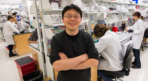
“The [CRISPR] technology demonstrates potential for many healthcare applications, including diagnosing infections in patients and detecting mutations that confer drug resistance or cause cancer,” stated senior author Feng Zhang, PhD. Zhang, shown above in the MIT lab named after him, is a Core Institute Member of the Broad Institute, Associate Professor in the departments of Brain and Cognitive Sciences and Biological Engineering at MIT, and a pioneer in the development of CRISPR gene-editing tools. (Photo copyright: MIT.)
Another unique use of CRISPR technology involved researchers David Liu, PhD, and Weixin Tang, PhD, of Harvard University and Howard Hughes Medical Institute (HHMI). Working in the Feng Zhang laboratory at the Broad Institute, they developed a sort of “data recorder” that records events as CRISPR-Cas9 is used to remove portions of a cell’s DNA.
They published the results of their development of CRISPR-mediated analog multi-event recording apparatus (CAMERA) systems, in Science. The story was also covered by STAT.
“The order of stimuli can be recorded through an overlapping guide RNA design and memories can be erased and re-recorded over multiple cycles,” the researchers noted. “CAMERA systems serve as ‘cell data recorders’ that write a history of endogenous or exogenous signaling events into permanent DNA sequence modifications in living cells.”
This creates a system much like the “black box” recorders in aircraft. However, using Cas9, data is recorded at the cellular level. “There are a lot of questions in cell biology where you’d like to know a cell’s history,” Liu told STAT.
While researchers acknowledge that any medical applications are in the far future, the technology holds the potential to capture and replay activity on the cellular level—a potentially powerful tool for oncologists, pathologists, and other medical specialists.
Using CRISPR to Detect Viruses and Infectious Diseases
Another recently developed technology—DNA Endonuclease Targeted CRISPR Trans Reporter (DETECTR)—shows even greater promise for utility to anatomic pathology groups and clinical laboratories.
Also recently debuted in Science, the DETECTR system is a product of Jennifer Doudna, PhD, and a team of researchers at the University of California Berkeley and HHMI. It uses CRISPR-Cas12a’s indiscriminate single-stranded DNA cleaving as a biosensor to detect different human papillomaviruses (HPVs). Once detected, it signals to indicate the presence of HPV in human cells.
Despite the current focus on HPVs, the researchers told Gizmodo they believe the same methods could identify other viral or bacterial infections, detect cancer biomarkers, and uncover chromosomal abnormalities.
Future Impact on Clinical Laboratories of CRISPR-based Diagnostics
Each of these new methods highlights the abilities of CRISPR both as a data generation tool and a biosensor. While still in the research phases, they offer yet another possibility of improving efficiency, targeting specific diseases and pathogens, and creating new assays and diagnostics to expand medical laboratory testing menus and power the precision medicine treatments of the future.
As CRISPR-based diagnostics mature, medical laboratory directors might find that new capabilities and assays featuring these technologies offer new avenues for remaining competitive and maintaining margins.
However, as SHERLOCK demonstrates, it also highlights the push for tests that produce results with high-specificity, but which do not require specialized medical laboratory training and expensive hardware to read. Similar approaches could power the next generation of POC tests, which certainly would affect the volume, and therefore the revenue, of independent clinical laboratories and hospital/health system core laboratories.
—Jon Stone
Related Information:
Multiplexed and Portable Nucleic Acid Detection Platform with Cas13, Cas12a, and Csm6
Rewritable Multi-Event Analog Recording in Bacterial and Mammalian Cells
CRISPR-Cas12a Target Binding Unleashes Indiscriminate Single-Stranded DNase Activity
Researchers Advance CRISPR-Based Tool for Diagnosing Disease
CRISPR Isn’t Just for Gene Editing Anymore
CRISPR’s Pioneers Find a Way to Use It as a Glowing Virus Detector
With New CRISPR Inventions, Its Pioneers Say, You Ain’t Seen Nothin’ Yet
New CRISPR Tools Can Detect Infections Like HPV, Dengue, and Zika
Breakthrough DNA Editing Tool May Help Pathologists Develop New Diagnostic Approaches to Identify and Treat the Underlying Causes of Diseases at the Genetic Level
CRISPR-Related Tool Set to Fundamentally Change Clinical Laboratory Diagnostics, Especially in Rural and Remote Locations
Harvard Researchers Demonstrate a New Method to Deliver Gene-editing Proteins into Cells: Possibly Creating a New Diagnostic Opportunity for Pathologists
Feb 23, 2018 | Instruments & Equipment, Laboratory Instruments & Laboratory Equipment, Laboratory Management and Operations, Laboratory News, Laboratory Operations, Laboratory Pathology, Laboratory Testing, Management & Operations
Researchers successfully isolated both plant and human RNA and DNA in the field, demonstrating the potential for their new dipstick technology to identify deadly bacteria, pathogens, and diseases in water, food, and even humans
Australian researchers at the University of Queensland (UQ) have developed an intriguing “dipstick” technology that might make it possible to use simple equipment to sequence DNA and RNA in the field. Among the potential applications that will interest clinical laboratory professionals is the ability for this technology to identify pathogens, both in humans and the environment.
Medical laboratories and anatomic pathologists are aware that gene sequencing (AKA, Nucleic Acid Sequencing) is the coming revolution in diagnostics. But the process is still costly and anchored to immovable technology that requires controlled environments and reliable resources. This promising new technology could make it simpler, cheaper, and faster to extract human DNA and RNA in settings outside a sophisticated core medical laboratory.
The UQ researchers developed technology that could affect how and where diagnostic tests for a whole range of pathogens are performed. For example, tests for bacteria such as E. coli in water supplies, pathogens in food, and diseases in humans currently are conducted in environmental and clinical laboratories. This new technology may allow such diagnostics to be done in extremely remote environments.
Isolating DNA/RNA in the Field
Jimmy Botella, PhD, Professor of Plant Biotechnology, and Michael Mason, PhD, Senior Post-doctoral researcher, both at the University of Queensland, led a team of researchers who published their findings in the journal PLOS Biology. The team developed a process they called “dipstick technology,” which allows DNA and RNA to be isolated quickly and without the use of specialized equipment.
They began by using the technology on particular plants, but soon found it could be used in many other situations.
“We found it had much broader implications as it could be used to purify DNA or RNA from human blood, viruses, fungi, and bacterial pathogens from infected plants or animals,” Botella noted in a press release.
The researchers’ objective was to investigate whether or not several different materials could be used to extract nucleic acids. “The first step in any application aiming to amplify DNA or RNA is the extraction of nucleic acids from a complex biological sample; a task traditionally requiring specialized equipment, trained technicians, and multiple liquid handling steps,” they wrote in the published study.

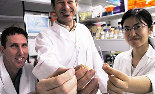
Holding the dipstick technology (from left) Dr. Michael Mason, Professor Jimmy Botella, and Yiping Zhou, all researchers at the University of Queensland in Brisbane, Australia. (Caption and photo copyright: University of Queensland.)
Their aim was to find a simpler process that required far less personnel and equipment. They found that cellulose-based filter paper could be used to bind nucleic acids. The filter paper, which was the control early in their investigation, even retained the nucleic acids through a purification process that removed contaminants. “We then adapted the cellulose filter to create a dipstick that can be used to purify nucleic acids from a wide range of plant, animal, and microbe samples in less than 30 seconds without the need for specialized equipment,” the researchers reported.
The team conducted its first tests on the plant species A. thaliana, a flowering plant found in Africa and Eurasia. However, wanting their dipstick technology to be useful in the field, they expanded their experiments to include various species of wheat, rice, soybean, tomato, and other plants. Citrus plants, known to be challenging, also were successfully tested.
The researchers then tested if their new technology would be useful for applications in humans, which is more complicated. HIV and hepatitis can be diagnosed using commercial kits, but those kits are not useful in many settings because the samples often require sophisticated manipulation. The researchers’ method—using cellulose paper and a one-minute wash—succeeded in amplification of the nucleic acid.
Performing Diagnostics in Hospitals, on Farms, and Even in the Jungle!
The University of Queensland’s commercialization company, UniQuest, has filed a patent application for the new technology. They are currently seeking partners to commercialize and sell the dipstick technology worldwide.
“Our dipsticks, combined with other technologies developed by our group, mean the entire diagnostic process from sample collection to final result could be easily performed in a hospital, farm, hotel room, or even a remote area such as a tropical jungle,” Botella noted in the press release.
The team conducted much of their field research on remote plantations in Papua New Guinea. They conducted tests on trees, livestock, human diseases, and to detect pathogens in food and water. “The dipstick technology makes diagnostics accessible to everyone,” Botella told Technology Networks.
Dipstick Diagnostics Not New to Point-of-Care Testing
As Modern Healthcare Executive noted, dipstick technology for various diagnostic purposes is not new, even though this particular application is, potentially, revolutionary. There are dipstick tests for everything from pregnancy to cholera. Also referred to as point-of-care testing (POCT), research and development of this technology has steadily grown, and as the UQ study shows, will likely continue.
In a paper published in Clinical Biochemistry Reviews, authors Andrew St. John, PhD, of ARC Consulting, and Christopher Price, MD, of the University of Oxford, noted, “Healthcare is changing, partly as a result of economic pressures, and also because of the general recognition that care needs to be less fragmented and more patient-centered.”
While there are certainly advantages to quick diagnostic tests that can be conducted in the field, there are some challenges, as well. Julie L. V. Shaw, PhD, Assistant Professor, Department of Pathology and Laboratory Medicine at The University of Ottawa, argues that “there are many challenges associated with POCT, mainly related to quality assurance,” in a paper she published in the journal Practical Laboratory Testing.
Technology will continue to develop and drive innovation and change in how diagnostics are performed and thus in how clinical laboratories operate. Various initiatives driving the industry toward personalized medicine and value-based care are sure to play a role, alongside new technology and other advancements.
With all of those changes, one thing remains critically important and that is the value of human understanding and innovation.
—Jillia Schlingman
Related Information:
Nucleic Acid Purification from Plants, Animals and Microbes in Under 30 Seconds
UQ Dipstick Technology Could Revolutionize Disease Diagnosis
Dipstick Technology Enables Rapid Diagnosis Anywhere
Existing and Emerging Technologies for Point-of-Care Testing
Practical Challenges Related to Point of Care Testing
The March of Technology Through the Clinical Laboratory and Beyond
Jan 3, 2018 | Laboratory Instruments & Laboratory Equipment, Laboratory News, Laboratory Operations, Laboratory Pathology, Laboratory Testing
University of Turin study in Italy shows under-vacuum sealing systems reduce exposure to formaldehyde by 75% among nurses handling tissue biopsy specimens during surgery
Histology technicians and anatomic pathology (AP) laboratories regularly handle dangerous chemicals such as formaldehyde. They understand the risks exposure brings and take precautions to minimize those risks. However, in operating suites worldwide, nurses assisting surgeons also are being exposed to this nasty chemical.
Nurses must place biopsies and other tissues into buckets of formaldehyde to preserve the tissue between the operating room (OR) and histology laboratory. Formaldehyde, along with toluene, and xylene, is used to process and preserve biopsy tissue, displace water, and to create glass slides. It is an important substance that has long been used to maintain the viability of tissue specimens. Thus, exposure to formaldehyde among nurses is well-documented.
According to a National Academy of Sciences report, formalin, a tissue preservative that is a form of formaldehyde, has been linked to:
· Myeloid leukemia;
· Nasopharyngeal cancer; and,
· Sinonasal cancer.
However, as Dark Daily previously reported, “One alternative to storing specimens in buckets with formalin is to vacuum-seal specimens … [so] that both the quality management of the patient specimen and worker safety for handling the specimens are greatly improved.” (See Dark Daily, “Anatomic Pathology Labs Adopt New Ways to Package, Transport, and Store Specimens to Reduce Formalin and Improve Staff Safety in Operating Theaters and Histology Laboratories,” October 13, 2014.)
Now, motivated by increasing formaldehyde regulations in Europe, as well as the need to increase awareness of exposure risks, the University of Turin (Unito), and other hospitals in Italy’s Piedmont region, conducted a cross-sectional study of 94 female nurses who were being potentially exposed to formaldehyde.
Researchers Aim for “Formalin-Free” Hospitals
The Unito study showed that nurses using an under-vacuum sealing (UVS) system in ORs are exposed to levels of formaldehyde 75% lower than those who did not use the system. This study differs from other similar tests because the level of exposure is not just potential, due to environmental contamination, but confirmed with analytic data from specific urine analyses.
The researchers divided the nurses into two groups:
· One group immersed samples in containers of formaldehyde following standard procedures;
· The other group worked in operating rooms equipped with a UVS system.
The researchers described a UVS system that called for the tissue removed during surgery to be sealed in a medical grade vacuum bag and refrigerated at four degrees centigrade before being transferred to the lab for fixation.
One example of a UVS system is TissueSAFE plus, developed by Milestone Medical, located in Bergamo, Italy, and Kalamazoo, Mich. According to the company’s website, the system, “Eliminates formalin in the operating theatre and allows a controlled formalin-free transfer of biospecimens to the laboratory.”

The image above is from a research paper by Richard J. Zarbo, MD, Pathology and Laboratory Medicine, Henry Ford Health System. It describes “five validation trials of new vacuum sealing technologies that change the approach to the preanalytic ‘front end’ of specimen transport, handling, and processing, and illustrate their adaptation and integration into existing Lean laboratory operations with reduction in formalin use and personnel exposure to this toxic and potentially carcinogenic fixative.” (Image copyright: Henry Ford Health System/Springer International Publishing.)
Increased Scrutiny Leads to New Pathology Guidelines
In a paper published in Toxicology Research, a journal of The Royal Society of Chemistry, the researchers noted a marked difference related to the adoption of the under-vacuum sealing procedure, as an alternative to formaldehyde for preserving tissues. “Nurses, operating in surgical theatres, are traditionally exposed to formaldehyde because of the common and traditional practice of immersing surgical samples, of a size ranging between two and 30 centimeters, in this preservative liquid (three to five liters at a time) to be later transferred to a [histopathology] lab,” the authors wrote. “We evaluated the conditions favoring the risk of exposure to this toxic reagent and the effect of measures to prevent it.”
Throughout Europe, increased scrutiny has forced medical pathology associations to write new guidelines that accept alternative methods to formaldehyde-based tissue preservation methods.
“In Europe, and in Italy in particular, the level of attention to formaldehyde exposure in the public health hospital system has become very high, forcing pathology associations to rewrite guidelines,” Marco Bellini, General Manager of the Medical Division at Milestone Medical, told Dark Daily. “What makes this study unique from many other similar tests is that the level of exposure has been confirmed with data from specific urine analyses,” he added.
The Italian Society of Pathological Anatomy and Diagnostic Cytology (SIAPEC), a division of the International Academy of Pathology, wrote general guidelines for AP labs that have been accepted and officially published by the Italian Ministry of Health.
The main topic of these guidelines is the preanalytical aspects of specimen collection, transportation, and preservation, where the vacuum method has been indicated as a valid alternative to improve the standardization of these crucial steps in pathology. By moving the starting point for specimen fixation from the OR to the histology labs, parameters can be controlled and documented, with the main advantage of reducing formaldehyde exposure by operators at the collection point.
These guidelines will be presented at the European Society of Pathology (ESP) with the intent to extending them throughout Europe.
Toluene’s and Xylene’s Effects Studied
Formaldehyde is not the only potentially harmful substance in the clinical laboratory. As previously noted, common solvents toluene and xylene also are potentially hazardous.
In fact, a study of pathologists, lab technicians, and scientists who work with toluene and xylene published in the Journal of Rheumatology found that the chance of acquiring Raynaud Syndrome (a vascular condition) doubled for those workers. (See Dark Daily, “Health of Pathology Laboratory Technicians at Risk from Common Solvents like Xylene and Toluene,” July 5, 2011.)
Medical laboratory leaders are reminded to initiate processes that ensure safe specimen handling, transport, and processing, as well as workflow changes that eliminate chemical odors in the lab. Studies, such as those cited above, may provide information necessary to affect change.
—Donna Marie Pocius
Related Information:
Formaldehyde Fact Sheet
Towards a Formalin-Free Hospital: Levels of 15-F2t-isoprostane and malondialdehyde to Monitor Exposure to Formaldehyde in Nurses from Operating Theatres
Histologic Validation of Vacuum Sealed, Formalin-Free Tissue Preservation, and Transport System
Notes Regarding the Use of Formalin, Reclassified as “Carcinogenic”
Formaldehyde Substitute Fixatives: Analysis of Macroscopy, Morpholologic Analysis, and Immunohistochemical Analysis
Anatomic Pathology Labs Adopt New Ways to Package, Transport, and Store Specimens to Reduce Formalin and Improve Staff Safety in Operating Theaters and Histology Laboratories
Health of Pathology and Laboratory Technicians at Risk from Common Solvents Like Xylene and Toluene
National Academy of Sciences Confirms that Formaldehyde Can Cause Cancer in a Finding that has Implications for Anatomic Pathology and Histology Laboratories


