New Test Under Development That Detects Breast Cancer within One Hour with 100% Accuracy Has Potential to Help Pathologists Deliver More Value
Such a test, if proved safe and accurate for clinical use, could be a useful diagnostic tool for anatomic pathologists
What would it mean to anatomic pathology if breast cancer could be diagnosed in an hour from a fine needle aspiration (FNA) rather than a core biopsy? A new test created by researchers affiliated with Massachusetts General Hospital in Boston may be just such a game changer. Especially in remote locations where clinical laboratory resources are in short supply.
Regardless of how the next round of research and clinical studies turn out, one reason this development is significant is that it demonstrates how newer technologies and analytical software are being combined to create a faster diagnostic test for different types of cancer.
Another benefit to this research is that it may utilize simpler, less expensive instruments. In fact, the researchers said this test can be performed for about $5. For these reasons, pathologists may want to follow the progress of these researchers as they work to improve this test so it can be used in clinical care.
Affordable Image Cytometry of FNA Specimens
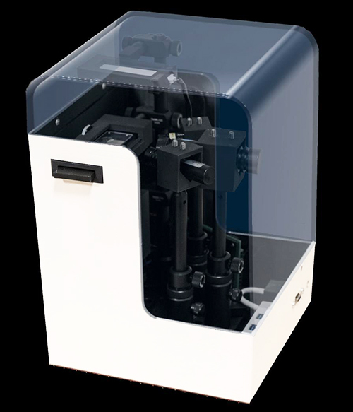
Though still in development, the new image cytometry system, dubbed CytoPAN, has demonstrated the ability to diagnose breast cancer within a one-hour time frame, and, according to the study published in Science Translational Medicine, “is devoid of moving parts for stable operations, harnesses optimized antibody kits for multiplexed analysis, and offers a user-friendly interface with automated analysis for rapid diagnoses.”
The international researcher team included scientists from:
- Massachusetts General Hospital (MGH),
- Nanyang Technological University, Singapore,
- Harvard-Massachusetts Institute of Technology (MIT), and
- Kyungpook National University, South Korea.
“Here, we report the development and validation of an affordable image cytometry system that allows automated and same-day molecular analyses of fine needle aspiration (FNA) specimens. Termed CytoPAN, for portable fluorescence-based image cytometry analyzer, the system performs multichannel imaging for cancer diagnosis and subtyping,” the researchers wrote.
The CytoPAN technique is minimally invasive, they note, and only requires a few cellular specimens to determine if breast cancer cells are present, with results available in one hour.

“Unfortunately, in many low- and middle-income countries, [breast cancer] diagnosis often takes an extraordinarily long time—up to a few months—due to a lack of specialists and limited laboratory infrastructure,” Hyungsoon Im, PhD, Assistant Professor at Harvard Medical School and one of the researchers involved in the project, told United Press International (UPI).
“From a public health aspect, it is critically important to develop new diagnostic methods that overcome these barriers,” he added.
Because FNA testing is less invasive than surgical biopsy collection, it has fewer complications and is generally considered safe. Thus, it is “feasible to be performed even in resource-limiting settings at much lower costs,” Im told UPI. “This could lead to earlier treatment and accelerate new drug testing in clinical trials.”
CytoPAN Testing and Additional Trials
The researchers tested CytoPAN on 68 breast cancer patients in South Korea.
“To determine the clinical utility of the approach,” they wrote in the published study, “we next conducted a prospective clinical study in which the FNA could be directly compared to conventional pathology results. We enrolled treatment-native patients at the Kyungpook National University Chilgok Hospital (Daegu, South Korea) and who were referred for primary surgery. All patients consented to have a preoperative breast FNA before clinically indicated surgery. The breast masses were visualized by ultrasound or computed tomography, and a coaxial needle was introduced through which FNA samples (CytoPAN) and core biopsies were obtained. Surgical specimens and/or core biopsies were processed by routine pathology and served as the gold standard.”
The CytoPAN platform detected the presence of breast cancer cells with a 100% accuracy, using as few as 50 harvested cells per collected specimen.
The test also successfully identified two key breast cancer biomarkers:
- hormonal receptors (ER/PR) with an accuracy rate of 93%, and
- epidermal growth factor receptor two with an accuracy rate of 96%.
“We are also preparing additional trials in the US and other countries,” Im told UPI. “The success in those trials will (hopefully) accelerate … widespread adoption of the technology.”
The researchers are currently testing CytoPAN on a larger number of patients in Botswana, with funding from the US federal National Institutes of Health (NIH).
According to the American Cancer Society (ACS), approximately 300,000 individuals are diagnosed with breast cancer annually in the US. The Union for International Cancer Control (UICC) states on their website that, globally, there are more than two million new cases of breast cancer diagnosed each year. And more than 600,000 people died from breast cancer worldwide in 2018. A disproportionate number of those deaths occurred in developing countries that have limited resources to diagnose and treat the disease.
Additional Research for Other Applications in Cancer Testing and Pathology
The new CytoPAN technology requires minimal training, according to the researchers, and only costs about $5 per test kit. This is substantially less expensive than the price associated with other tests available on the market, UPI noted.
Though additional research and clinical trials are needed before CytoPAN will be available for widespread clinical use, a cost-effective, relatively non-invasive test that can accurately diagnose cancer within an hour would be transformational for anatomic pathology and, potentially, could save many lives.
—JP Schlingman
Related Information:
CytoPAN—Portable Cellular Analyses for Rapid Point-of-care Cancer Diagnosis
System Provides ‘Faster, Less Invasive’ Method for Breast Cancer Detection


